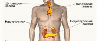
The pineal gland is an organ in the brain that functions as the endocrine system.
As is known, the functions of this gland have not yet been thoroughly studied due to its small size and location.
What is clearly known is that the gland regulates the processes of sleep and wakefulness in the body.
In some cases, the pineal gland becomes inflamed. What is the reason for this condition and why it is dangerous, we will look into this article.
Causes and symptoms of the disease
The main reasons for the appearance of pineal gland cysts are:
- Problems with the outflow of melatonin produced by the gland, which causes secretion to accumulate.
- Echinococcosis is a type of helminthiasis that causes cysts in various organs of the human body. To put it another way, the pineal gland is infected with the help of echinococcus, which enters the brain through the main bloodstream. The parasite forms a cocoon around itself, with the help of which it is protected from the body’s immune system. The cyst is the shell of the parasite itself, which is gradually filled with the products of its vital activity and can slightly increase in size.
These are the main reasons that have been proven by medicine, but there may be others. Since the disease occurs with virtually no symptoms, other causes cannot yet be determined.
As for the symptoms of a pineal gland cyst, they include:
- Constant headaches that occur for no apparent reason
- Double vision, blurred vision and other visual impairments
- Possible disturbances in the clarity of human movements and gait
- Hydrocephalus
- Persistent state of depression, dementia
- Hypertension
Symptoms depend entirely on the volume of the cyst and the stage of the disease. So, if the cyst is small, then symptoms may hardly appear at all.
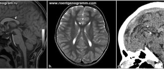
MRI in the diagnosis of pineal gland pathologies
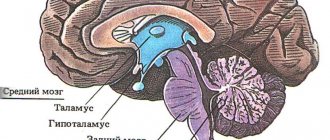
The epiphysis (pineal or pineal gland) is a gland located between the anterior tubercles of the quadrigeminal plate, connected through a stalk to the third ventricle.
Among pineal gland diseases detected by MRI examination, manifested by certain clinical symptoms and asymptomatic, the majority are tumors of the pineal region.
There are several types of neoplasms affecting the pineal gland:
Tumors arising from the parenchyma of the pineal gland (pineocytomas, pineoblastomas)
Pathological processes occurring in the pineal gland, in most cases, are manifested by premature puberty, which is an indication of the endocrine function of this organ.
Pineocytoma. On T2 WI, the tumor consists of two areas of different MR signal intensity: high in the anterior parts and low in the posterior parts. The clarity and linearity of the transition indicates the cystic nature of the tumor structure. Differences in the intensity of the MR signal from the fluid in the anterior and posterior parts of the tumor are due to the effect of “sedimentation” of protein components (most likely blood components), moving down when the patient’s head is positioned on the back of the head.
Pineocytoma. MRI in T2 and T1 mode reveals a solid tumor of the posterior parts of the third ventricle of the brain. Against the background of IV contrast, a pronounced and fairly homogeneous nature of tumor contrast is determined.
Pineoblastoma. In T2 and T1 mode, a tumor of the posterior parts of the third ventricle of the brain with the development of occlusive hydrocephalus is detected. There is a small area of subacute hemorrhage in the tumor stroma. After IV contrast, the tumor intensively and fairly homogeneously accumulates the contrast agent.
Germ cell tumors of the pineal gland (germinoma, teratoma, mixed germ cell tumors)
These formations develop from germinal cells, which during embryogenesis did not reach their normal location in the gonads and are similar in structure and function to tumors arising from cells of the testes and ovaries.
Germ cell tumors grow into the walls of the third ventricle and the hypothalamus, which determines the typical triad of symptoms: hypogonadism, optic nerve atrophy and diabetes insipidus.
In addition, the tumor can compress the cerebral aqueduct, which leads to the development of occlusive hydrocephalus, manifested by severe headaches, vomiting, impaired consciousness, and swelling of the optic nerve nipple.
Compression of the quadrigeminal tubercles may cause Parinaud's syndrome (combined upward gaze palsy), and pressure on the cerebellum or brainstem may result in gait disturbance.
Germinoma of the pineal region. On T2 WI the tumor has a weakly hyperintense signal compared to the brain tissue, while on T1 WI the tumor is almost isointense with the brain. The ventricular system is hydrocephalically dilated. The third ventricle is deformed. The quadrigeminal plate is pushed posteriorly.
Germinoma of the pineal region. The tumor has a heterogeneous structure with many small and large cysts. Perifocal edema is detected around the tumor.
Sometimes hypothalamic symptoms come to the fore in the clinical picture: hyperphagia, anorexia, impaired thermoregulation.
Germ cell tumors of the pineal gland can metastasize to other areas. In some cases, germinomas spread to the area of the sella turcica, manifesting itself with the symptoms of pituitary tumors.
Germinoma of the pineal region with metastasis to the chiasmal-sellar region.
Germinoma of subcortical formations on the left. A multicystic tumor with signs of occlusion of the foramen of Monroe on the left.
Malignant mixed germ cell tumor. In the lateral ventricles and pineal region, multiple tumor nodes with pronounced peritumoral edema are detected. Intraventricular formations with signs of hemorrhage. With intravenous contrast, pronounced contrasting of the nodes is determined. Additionally, metastasis is visualized along the ependyma of the lateral and fourth ventricles of the brain.
Glial tumors of the pineal gland (astrocytoma, ependymoma, glioblastoma, etc.)
This type of tumor originates from glial cells of neighboring brain structures, such as the thalamus, midbrain, quadrigeminal plate, splenium of the corpus callosum, with subsequent spread to the pineal region.
Pilloid astrocytoma of the posterior parts of the third ventricle. A small formation with the development of occlusive hydrocephalus is determined. The tumor practically does not differ in signals from the substance of the brain. After IV contrast, an increase in the MR signal from the formation is observed, which makes it possible to clarify the location of the tumor, the degree of compression of the quadrigeminal plate and the cerebral aqueduct.
Glioblastoma of the optic thalamus on the right. Before and after IV contrast, a space-occupying formation with a necrotic center and a contrasted peripheral infiltrative part is determined. The posterior sections of the third ventricle are deformed, the initial manifestations of occlusive hydrocephalus.
Ependymoma of the posterior parts of the third ventricle. A mass formation is detected in the posterior parts of the pineal gland. The anterior tubercles of the quadrigeminal are pushed down. The cerebral aqueduct is compressed.
Mixed tumors and non-tumor formations.
Dermoid tumor of the pineal region.
Lipoma of the pineal region.
Cavernoma of the pineal region.
Meningioma of the pineal region.
Epiphysis cyst. Compression of the quadrigeminal plate and partial compression of the cerebral aqueduct.
Currently, MRI is the main diagnostic method that allows us to give a fairly complete description of the pathological process located in the pineal region.
Multi-projection research - obtaining images in different planes and three-dimensional images (3D) help determine the location of the tumor in relation to the third ventricle and surrounding tissues, the predominant direction of growth, the relationship with the venous and other tissue formations of the pineal region, the degree of invasion of the adjacent medulla.
Treatment of the disease
Pineal gland cysts in most cases do not require medical treatment and cannot be treated.
Cysts caused by the presence of a parasite in the brain can be treated with medication only if the patient applies at the initial stage, which happens extremely rarely. In most cases, surgery will be required to solve the problem.
Direct indications for surgical intervention include the following factors:
- Clearly expressed symptoms that interfere with a person’s normal functioning.
- With a significant risk of developing hydrocephalus.
- If the cyst significantly affects the functioning of the cardiovascular system or affects surrounding brain structures.
Since the disease has not yet been fully studied, the factors causing the disease are unknown. Of course, surgery carries a certain risk to a person’s health and life, but in most situations, this is the only way out that will help get rid of the disease.
If the case is not critical, constant observation and monitoring of the patient's condition is allowed. In this case, MRI should be done at least 2 times a year. After an MRI, you need to consult a doctor who will compare the result with the previous one and monitor the dynamics of the development of the disease.
As for drug treatment, most often it is the following system of drugs:
- Adaptogens, which help the body align its wakefulness and rest cycles
- Preparations containing melatonin, a deficiency of which occurs during illness
- Diuretics and venotonics, which will help improve the outflow of fluid from the brain; drugs are prescribed if there is a risk of cerebral hydrocephalus
- If a headache is present, analgesics are often prescribed to relieve the symptom.
- Special medications are also prescribed to eliminate nausea and vomiting.
MRI is an effective method for diagnosing the disease
If the disease is at a serious stage and epilepsy attacks appear among the symptoms, the doctor selects medications to eliminate or alleviate them.
4. Treatment of tumors and cysts of the pineal gland
The main method of treating tumors is surgery
.
Most often, a neurosurgeon is faced with the need to choose a complex treatment method (a combination of radiation, surgical and drug therapy)
. The main difficulty of treatment is associated with the histological diversity of neoplasms and their dangerous proximity to large blood vessels.
As for cysts, in most cases they cannot be removed - they are under constant medical supervision
. The exception is when the cysts reach a large size and are removed through surgery.
If hydrocephalus develops, drainage operations are performed to ensure the outflow of excess fluid.
Throat spasm: causes and methods of treating pathology
Discussion: 3 comments
- Elena:
07/02/2017 at 13:57During an MRI of the brain, I was diagnosed with a pineal gland cyst. My symptoms include infrequent headaches and sleep problems. Every day I have difficulty falling asleep, my sleep is superficial and I have frequent dreams. The doctor prescribed Melaxen 10 minutes in advance. before sleep.
Dmitry Ivanov:
11/21/2019 at 09:56
Melaxen, like melatonin-sz, are excellent drugs that help restore normal sleep, and the quality of sleep itself increases. What I also like is that they are not prescription drugs, buying them is not a problem.
Elya:
12/16/2019 at 14:48
Adaptogens are generally a cool thing; I myself take melatonin-sz from time to time. Very pleased!
Magnetic resonance imaging of epiphysis calcifications
The etiological factors of excess calcium deposition in the pineal gland tissue have not been reliably established. To identify calcifications, MRI is not necessary, since dense structures are shown by CT. Computed tomography is accompanied by radiation exposure of tissue, so indications for the method are strictly limited. When using radiation diagnostic methods in order to reduce radiation exposure, it is enough to do a targeted examination with a limitation of the area of exposure.
Calcification forms at the site of hemispheric defects. Pathological changes develop at the sites of impaired blood supply, inflammatory, and infectious lesions. At the beginning of the development of nosology, the normal dimensions of the epiphysis on MRI change, and areas of altered density appear. To detect defects at the beginning of formation, it is rational to do tomography with high-field devices with the ability to identify pathological foci with a diameter of 1 mm.
Types of pineal gland calcification
The prevalence of physiological calcifications is observed in people after 50 years of age. Areas of increased calcium accumulation are localized in the cerebellum, choroid plexus, falx, epiphysis, and arachnoid granulation. Calcification of the nerve ganglia in young people under thirty years of age is a sign of pathological processes.
Dystrophic calcification occurs against the background of chronic infections and long-term persistence of epidural hematoma. There are practical facts of the appearance of calcifications after chemotherapy, radiation exposure, leukoencephalopathy, microangiopathy, and degenerative diseases in the elderly.
Congenital calcification in children is formed due to neurofibromatosis, basal cell nevus syndrome.
Giant cell astrocytomas and areas of sclerosis also often undergo calcification.
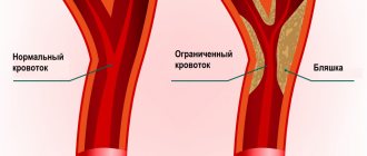
Vascular calcifications appear with atherosclerosis, malformations, and aneurysms. Typical localization is the middle, anterior cerebral artery.
Congenital infections with calcium deposition in the pineal gland - rubella, toxoplasmosis, cytomegalovirus, herpes simplex, HIV, tuberculosis, cysticercosis, sarcoidosis.
Tumors with calcification:
- Meningiomas;
- Craniopharyngiomas;
- Ependymomas;
- Astrocytomas;
- Oligodendrogliomas;
- Glioblastoma.
Metastatic foci of brain damage provoke calcification of the pineal gland.
The accumulation of lime rarely leads to disruption of cerebral functionality. Atherosclerotic plaques limit the patency of the vessel, which causes the development of hypoxia and stroke.
Calcifications can contribute to hormonal imbalances. Hypoxic conditions disrupt the functionality of the basal ganglia, hypothalamus, and pituitary gland. Progression of the condition leads to hypothyroidism, damage to thyroid function.
Preventive measures
Due to insufficient research on the pineal gland, there are no specific preventive measures. In general, to prevent the development of cystic formation in the gland, it is necessary to adhere to a healthy lifestyle, strengthen the body’s defenses (adequate physical activity, proper rest, emotional stability). It is necessary to adhere to a complete and balanced diet; it is recommended to consume foods with a high tryptophan content (fatty fish, oatmeal, nuts, protein products, cottage cheese).
Kinds
There are 5 types of cysts:
- arachnoid (the most common and most dangerous);
- colloidal;
- pineal;
- dermoid;
- epidermoid.
An arachnoid cyst of the pineal region or cerebrospinal fluid is localized in the arachnoid membranes of the brain, filled with CSF. More often diagnosed in men, it develops after inflammation and infections. Gives symptoms in case of increased ICP and compression of an area of the cerebral cortex with its increased growth.
Colloidal cysts are usually congenital and do not manifest themselves for a long time. If there are disturbances in liquorodynamics, they can cause the development of intracerebral hernias and hydrocephalus with a fatal outcome.
Dermoid and epidermoid cysts manifest themselves during embryogenesis. The contents of such cysts include fat, hair, etc.
They tend to grow rapidly, which indicates the need for urgent removal after the birth of the child.
Pineal cysts are neoplasms of the body of the gland; they are always very small and harmless.