The human brain is called the most mysterious and perfect creation of nature. It controls all functions of the body and ensures that a person carries out intelligent activities. Here, all information received from the external environment and internal environment of the body is analyzed and appropriate human behavior is formed. And if animals receive information from specific objects and phenomena, then for humans the word becomes the real signal. Word and speech constitute the second signaling system, unique to humans. The material substrate of the second signaling system and verbal human thinking is the cerebral cortex. Scientists have been trying to unravel the mysteries of the human brain for many centuries, but even now they are still very far from knowing the truth.
What is the brainstem and what are its similarities to the spinal cord?
Anatomy of the brainstem. The brain stem (BM) includes:
- Medulla,
- Pons,
- midbrain,
- Diencephalon.
GM trunk - located between the spinal cord and telencephalon. The cerebellum is closely connected to the brainstem through the peduncles.
Similarities between the GM and SM trunks (spinal cord):
- SM is the beginning of the spinal nerves. The GM trunk is the beginning of 11 pairs of CN (cranial nerves).
- Similar relative positions of gray and white matter.
Structure and functions of the cerebellum
The cerebellum is located above the brain stem and is connected to its parts by 3 pairs of peduncles. The cerebellum has 2 small hemispheres covered by the cerebellar cortex. The main functional significance of the cerebellum is to maintain body balance, regulate and coordinate body movements, giving them smoothness, accuracy and proportionality. The cerebellum programs the automatic execution of movements, which is made possible by its connections with the spinal cord, brainstem and cerebral cortex. For example, when walking and running, the cerebellum controls the position and movement of the torso and arms in accordance with the movements of the legs and the movement of the body's center of gravity. When writing, it is responsible for maintaining optimal posture and coordinating the movements of the head, eyes and hands. The cerebellum plays an important role in performing rapid sequential and simultaneous movements, such as the movements of the hands of a pianist or typist.
Differences between the brainstem and the spinal cord
How does the anatomy of the brainstem differ from the structure of the spinal cord?
1) SM - segmental structure. The GM trunk is not (innervation zone of the CN).
2) Gray matter SC - continues continuously. GM trunk - gray matter is divided into nuclei.
3) CM cavities - central canal. GM barrel cavities have different structures:
- 4th ventricle (tent shape), the bottom of the 4th ventricle is a rhomboid fossa.
- the midbrain is a narrow canal (plumbing).
- hindbrain - 3 ventricles (between the visual thalamus).
Structure and functions of the cerebrum
The right and left hemispheres form the so-called telencephalon, or cerebrum, which is the most developed and, in evolutionary terms, a new part of the brain. The work of the cerebral hemispheres is associated with the most complex manifestations of human mental and intellectual activity.
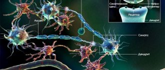
Gray and white matter of the brain The surface of the hemispheres is covered with the cerebral cortex - a layer of gray matter consisting of nerve cells (neurons). It is here that the highest analysis of all received information occurs and human behavior is formed. Under the cerebral cortex in the hemispheres there is white matter formed by the processes of neurons (nerve fibers). Bundles of nerve fibers form pathways that connect the cerebral cortex with other parts of the brain and with the spinal cord. The right and left hemispheres of the cerebrum are connected to each other by a huge number of nerve fibers, the totality of which is called the corpus callosum.
The significance of the basal ganglia In the depths of the white matter of the hemispheres there are accumulations of gray matter - the basal ganglia, which control the automated movements of the body, control and maintain the tone of skeletal muscles, and regulate their heat production. When connections between the basal ganglia and the motor centers of the midbrain are disrupted, parkinsonism develops, which is characterized by severe trembling of the limbs and head. One of the basal ganglia, the amygdala, is an important part of the limbic system of the brain. Its destruction leads to aggressive behavior or, conversely, a sluggish, apathetic state.
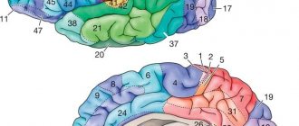
Convolutions and grooves of the brain The cerebral cortex forms folds - convolutions, which are separated by grooves. Due to this relief, the surface of the cerebral cortex increases. Deep grooves divide each hemisphere into lobes: frontal, parietal, occipital, temporal, limbic and insular. Smaller grooves within each lobe have an individual pattern and are formed in a person from birth to 7–8 years.
Motor Center Thanks to numerous clinical observations and scientific studies, it has been established that specific brain functions are associated with certain areas of the cortex. Based on the available data, at the beginning of the twentieth century, K. Brodman identified 52 fields of the cerebral cortex, and currently there are more than 200 of them.
According to modern concepts, the motor center is located in the frontal lobe, in the area of the precentral gyrus (on the border with the parietal lobe). Information from the muscles and joints of the body comes here, based on the analysis of which conscious regulation of movements is carried out. When this area of the cortex is damaged (for example, due to a stroke), paralysis of the muscles of the opposite half of the body occurs.
Writing Center and Speech Motor Center The frontal lobe contains the writing center and speech motor center. Defeat of the first leads to disorders of writing skills under visual control (agraphia). The speech motor center has a pronounced functional asymmetry: if it is disrupted in the right hemisphere, the ability to regulate timbre and intonation is lost (speech becomes monotonous); if it is destroyed on the left, the ability for articulate speech (aphasia) and singing (amusia) is lost. With partial disorders, agrammatism is possible - the inability to form phrases correctly. The location of other speech centers in the cortex is also asymmetrical: in right-handed people they develop in the left, in left-handed people - in the right hemisphere of the cerebrum.
Area of the frontal pole A large area of the cortex in the anterior part of the frontal lobe carries out programming of complex forms of behavior: planning actions, making decisions, analyzing the results obtained, volitional reinforcement. The area of the frontal pole is related to the control of a person’s psycho-emotional state. Damage to this area can affect a person’s character, his intellectual activity, value orientations and result in changes in personality structure.
Center of general sensitivity In the parietal lobe, in the postcentral gyrus, there is a center of general sensitivity (pain, temperature, tactile). Violations of the cortex in this area lead to partial or complete loss of sensitivity. Lesions of the cortex in other parts of the parietal lobe contribute to a disorder in the function of recognizing objects by touch, without the help of vision, as well as the ability to perform complex professional movements that require special training. In the area of the cortex of the parietal lobe, on the border with the temporal and occipital lobes, there is a visual (optical) speech center. When it is damaged, the ability to understand readable text is lost (Alexia).
Visual center In the occipital lobe, along the edges of the calcarine groove, the visual center is located. Its damage leads to blindness. If there are disturbances in the areas of the occipital lobe cortex adjacent to the calcarine sulcus, there may be a loss of visual memory, the ability to navigate in an unfamiliar environment, the ability to use vision to evaluate the shape of objects, the distance to them, and correctly measure movements in space.
Auditory center The auditory center is located in the middle part of the superior temporal gyrus. The consequence of its damage is deafness. Near it is the auditory speech center. Injuries in this area result in an inability to understand spoken language, which is perceived as noise. Other areas of the temporal lobe cortex are associated with the activity of the vestibular apparatus. If they are damaged, balance when standing is disrupted.
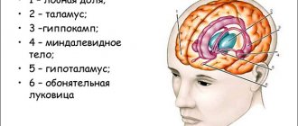
Limbic lobe The limbic lobe is located on the inner surface of the cerebral hemispheres facing each other. Its cortex controls a complex of functional and behavioral psycho-emotional reactions to environmental influences. The gustatory and olfactory centers are also located here. Associated with the limbic lobe, an area of the evolutionarily old cortex called the hippocampus plays an important role in human learning, as it influences memory mechanisms. The significance of the insular cortex is currently not well understood.
Differences between cranial nerves and spinal nerves: which ones are they divided into based on fiber composition?
SMN (spinal nerves) are mixed, CN are not all mixed.
According to the composition of CN fibers:
• 1, 2, 8 – only sensitive (nerves of the sense organs).
• 3, 4, 6, 11, 12 – motor fibers (similar to the anterior roots of the SC).
• 5, 7, 9, 10 – mixed.
• 3, 7, 9, 10 - have autonomic fibers - innervate the smooth muscles of internal organs, glands and cardiovascular system.
Causes of brainstem stroke
The following factors contribute to the development of the disease:
- Hypertonic disease. It is the most common cause of vascular damage to the brain stem. With high blood pressure, the walls of the arteries become brittle, which in turn leads to their rupture and hemorrhage.
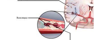
- Atherosclerosis, thrombosis. When the lumen of a vessel is blocked by an atherosclerotic plaque or thrombus, the blood supply to the surrounding tissue is disrupted, which leads to the development of ischemia.
- Aneurysms, collagenosis, vascular malformations. Pathological changes in the structure of the vascular wall increase the risk of its rupture. This anomaly can cause a stroke at a young age.
- Diabetes mellitus, rheumatological diseases, heart pathologies, blood clotting disorders, etc. Features of the course of some diseases lead to the development of atherosclerosis, blood clots, macro- and microangiopathy.
- Lifestyle. Smoking, alcohol abuse, physical inactivity, poor diet, stress, and chronic fatigue increase the risk of vascular accidents.
- Age. Each subsequent 10 years increases the likelihood of developing ischemia or hemorrhage by 5-8 times.
- Genetic predisposition. Cohort studies have revealed that the risk of stroke increases by 30% if a person has a history of this disease in their family.
Make an appointment
Patterns of location and projection of cranial nerve nuclei
The CN nuclei are located in the GM trunk.
- The nuclei of the last four (9-12) are in the medulla oblongata, the nerves exit from the medulla oblongata.
- The middle quadruple nuclei (5-8) are in the pons, the nerves exit the pons.
- Nuclei 3 and 4 pairs are in the midbrain, nerves emerge from the midbrain.
- 1 and 2 pairs of nuclei - there are no nodes, these are outgrowths of the brain (2 pairs - outgrowths of the diencephalon, 1 pair - outgrowths of the telencephalon into the nasal cavity; clinical significance - viruses and also medicines penetrate through them).
Projection of nuclei onto the rhomboid fossa.
The rhomboid fossa is the dorsal surface of the medulla oblongata and the pons. 8 pairs of CNs are projected onto it:
- Nuclei 9-12 pairs - on the lower half of the rhomboid fossa.
- Cores 5-8 pairs - on the upper half.
- 3 and 4 pairs - not related to the rhomboid fossa (in the midbrain).
Along the midline are projections of the motor nuclei . Laterally – projections of sensitive nuclei . Between them are vegetative nuclei .
Structure and functions of the diencephalon
Anterior to the brain stem, between the midbrain and the cerebral hemispheres, is the diencephalon. The upper part of the diencephalon is called the thalamus or thalamus optic, the lower part is the hypothalamus.
The meaning of the thalamus The thalamus, a paired ovoid-shaped formation, is a collector of all types of sensitivity from all parts of the body and sensory organs. From here this information is transmitted to the cerebral cortex. Certain areas of the thalamus are important components of the limbic system of the brain, which controls the psycho-emotional behavior of a person, while others are involved in ensuring memory processes. There is evidence that the thalamus is involved in the perception of pain. Destruction of certain areas of the thalamus can lead to a decrease in anxiety, tension, aggressiveness, elimination of obsessive thoughts, as well as a sharp decrease in motor activity.
The significance of the hypothalamus The significance of the hypothalamus is associated primarily with the regulation of the activity of internal organs. The nuclei of the hypothalamus produce special substances - neurohormones, which enter the pituitary gland, and from it into the blood.
The pituitary gland is an endocrine gland, closely related in structure and location to the hypothalamus. The single hypothalamic-pituitary system of the diencephalon controls the work of other endocrine glands and, with their help, regulates the functions of the body. This system controls the state of water-salt balance, metabolism and energy, the functioning of the immune system, thermoregulation, reproductive function of the body, etc. There is evidence that the hypothalamus contains specific pleasure centers that play an important role in the formation of motivations and emotional forms of behavior . In the area of the hypothalamus there are areas of the optic nerves through which information is transmitted from the retina of the eye.
The significance of the pineal gland The diencephalon also includes the pineal gland, or pineal gland, an endocrine gland that influences the work of other endocrine glands and is involved in the regulation of the seasonal rhythms of the body.
What is the medial loop, where is it formed, what is included in its composition and where does it end?
The medial lemniscus is a set of sensory pathways running through the lateral nucleus of the visual thalamus into the cortex.
Formed between the medulla oblongata and the pons.
The medial loop includes:
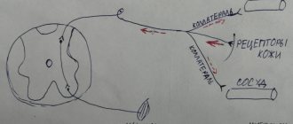
1) Spinothalamic tract (tractus spinothalamicus) - cutaneous sensation from the trunk and limbs.
2) Optic tubercular fasciculus - proprioceptive sense from the trunk and limbs.
3) Pathway - carries cutaneous and proprioceptive sensitivity from the head and neck (axons of neurons of sensory nuclei - 5,7,9,10 CN).
4) Vestibular pathway.
Brain topography
Each part of the brain has its own functions. For example, information obtained through vision
is analyzed in the occipital region of the brain.
And the movement
is controlled by a fairly narrow strip of nervous tissue that stretches from the top of the head to the ear, like the earpiece of headphones.
Brain training
Is your memory failing? It’s not surprising, because she suffers without load in the same way as her muscles. Memory development exercises will help you - you can start doing them at any age.
At the same time, vision, hearing, movement, and all tactile sensations are controlled in a mirror way.
So, if a person has a stroke in the left hemisphere, the motor functions of the right side of the body are impaired. Next to the motor area is the area where tactile sensations
.
Therefore, often when the brain is damaged, a person simultaneously loses both the ability to move and the ability to feel. The perception of auditory information
occurs in the temporal region of the brain.
In right-handed people, the left temporal lobe is responsible for understanding words and expressing one's own thoughts. Right temporal lobe – helps to hear music and identify various noises. The area of the brain where the visual and auditory areas meet is responsible for the function of reading
- converting visual images into sounds.
What tracts does the pyramidal tract at the trunk level divide into? Their purpose
Motor pathways are divided into: pyramidal and extrapyramidal.
The pyramidal tracts in the region of the GM trunk are divided into three tracts:
1) Tractus corticospinalis – motor activity of the muscles of the trunk and limbs (cortex => trunk => motor nuclei of the SC).
2) Tractus corticonuclearis – muscles of the head and neck (cortex => motor nuclei of the CN (3,4,5,6,7,9,10,11,12)).
3) Tractus corticopontocerebellaris (bark => trunk => cerebellum).
Interaction with other parts of the brain
The human central nervous system is a unique formation, under the control of which all internal systems of the body function, be it breathing or heartbeat.
An important role in this is played by the brain stem, which contains nuclei - the nerve centers of the corresponding structures.
With the help of them, the human body, at the subconscious level, carries out various reflexes that are under the control of the brain stem, and maintains the constancy of the internal environment, senses aromas, hears, sees and touches the world around us.
Types and consequences of stroke
There are two types of stroke: hemorrhagic and ischemic. Ischemic stroke is the most severe and life-threatening. Nowadays, ischemic stroke is the second leading cause of human mortality. Another name for this stroke is human brain infarction.
Ischemic stroke of the brain, as a result of which complete damage to the tissue particles of the brain occurs, and the cause of this disease is a circulatory disorder. Diseases that can cause ischemic stroke: various stages of atherosclerosis, diabetes mellitus of all types, hypertension and rheumatism of any degree of development. If you suddenly experience loss of coordination, dizziness, nausea. This indicates the development of a stroke.
Brain stem stroke can occur for a variety of reasons. First of all, a stroke occurs due to disruption of cerebral circulation. Unfortunately, a stroke almost always leads to death or severe damage to the brain stem, which is responsible for vital body functions.
A stroke occurs suddenly, dizziness, sweating, pallor, high temperature appear, blood pressure increases, and the pulse quickens. After this, problems with breathing and blood circulation arise, breathing becomes rapid and intermittent. Due to the disturbance of impulses, paralysis occurs, but mental activity does not suffer. A person is able to fully control the situation and think soberly.
With such a stroke, as a rule, two out of three people die, the crisis period is the first two days after the attack. Most often, death occurs during this period. The most important thing is to react quickly and seek help from doctors; within an hour from the moment of the attack you need to call an ambulance, then it is possible to save the person. Such a stroke is actually very difficult to treat; it requires treatment by qualified doctors and hospitalization.
In the most severe forms of stroke, there may even be surgery to prevent further bleeding, but such surgery is generally not performed. If you manage to survive after a stroke, then you will have a very long therapy, which can last more than one year. Depending on the severity of the stroke, rehabilitation is carried out at home, in recovery centers or special sanatoriums. It is impossible to survive a second stroke.
Diagnosis of brainstem stroke
Diagnosis of the disease begins with examination data and collection of the patient’s medical history. Based on the characteristic symptoms, a brain stem stroke can be suspected in the first minutes. Such patients are hospitalized on an emergency basis. In a hospital setting, a number of procedures are performed to confirm the diagnosis. The following have the greatest diagnostic capabilities:
- Computed tomography is an x-ray method for studying the layer-by-layer structure of organs and tissues. With a high probability it helps to recognize foci of hemorrhage in the brain.
- Magnetic resonance imaging is an effective method for detecting pathological lesions using nuclear magnetic resonance. Allows you to assess the degree and volume of tissue damage.
- Doppler ultrasound. Using ultrasound of the vessels of the neck and brain, pathological changes (stenosis, occlusion) can be detected in the carotid and vertebral arteries, as well as in their branches.
- Electroencephalography. It is also a non-invasive diagnostic method. By recording bioelectrical activity, it reflects qualitative and quantitative changes in the function of the cortex and deep structures of the brain.
In addition, it is possible to conduct laboratory tests of the composition of blood and cerebrospinal fluid. According to indications, a neurologist is able to draw up an almost complete clinical picture, which reflects the depth and localization of the damage that has occurred. But to confirm the diagnosis, additional studies are required using CT or MRI, which will show:
- type of disorder;
- the extent of the pathology;
- presence of hematoma or ischemia.
The more fully and accurately the factors of change are identified, the easier it is to draw up a rehabilitation plan.