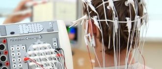
What is the essence of the EEG procedure?
An electroencephalogram records electrical signals from brain cells and allows us to identify epilepsy, trauma, neoplasms, inflammatory processes, and changes in blood vessels. Pathology is indicated by disturbances in the electrical activity of neurons, which are recorded using special sensors placed on the patient’s head.
Using an electroencephalogram, the doctor can:
- analyze the performance of the brain;
- identify foci of pathologies;
- assess the nature and extent of damage;
- confirm or clarify the diagnosis;
- monitor the effectiveness of the treatment.
In what cases is EEG prescribed?
After a conversation with the patient and studying the medical history, the specialist decides to prescribe encephalography. Typically, indications for an EEG are frequent headaches, sleep problems, fainting, fatigue and chronic fatigue.
Deterioration in well-being may be a sign of disorders in the functioning of the brain. The above ailments often arise due to:
- vegetative-vascular dystonia;
- cardiac dysfunction;
- pathologies of blood vessels of the neck and head;
- inflammatory processes in meningitis and encephalitis;
- endocrine disorders;
- malignant or benign neoplasms.
For patients suffering from epilepsy who have undergone neurosurgical interventions and head injuries, EEG monitoring is mandatory.
Prognosis for epilepsy.
Epilepsy is always assessed by a doctor as a complex of symptoms.
Benign forms of epilepsy often require only observation and may resolve on their own with age.
In other cases, epilepsy and associated pathologies may require serious treatment, and the prognosis will depend on the severity of the underlying disease. Some of these pathologies can be cured completely, for example, by removing a tumor or eliminating the consequences of an injury. Then the attacks will disappear. In other cases, lifelong medication and work with a psychologist are required.
How to prepare for an electroencephalograph examination?
Monitoring the electrical activity of the brain does not require complex training. During the examination, it is important to simply follow the doctor's instructions and remain calm. Flashes of light and noise are part of the procedure.
Three days before the examination, it is recommended to stop taking tranquilizers, anticonvulsants and sedatives. 24 hours before the examination, do not drink tea, coffee, energy drinks, or eat chocolate. The day before monitoring, wash your hair. Have a snack an hour before the examination. Before starting, loosen your hair and remove metal jewelry.
What should parents pay attention to?
The first and most important thing is to remember that epilepsy is not always seizures with loss of consciousness. In epilepsy, seizures can be local (local): twitching of the arm, leg, corner of the mouth, or they may be completely absent. Seizures can be manifested by blinking, drooling, redness of the face, episodes of “switching off” that the child does not remember, changes in behavior (the appearance of aggression, fear).
Often the child's status may only be developmental delay.
Not every seizure can be a manifestation of epilepsy that requires serious treatment. Convulsive syndrome is the body’s response to intoxication, endocrine diseases, fever, cerebrovascular accidents, increased intracranial pressure and other numerous pathologies.
Parents should be alert to:
- Asymmetry. Only one arm or leg, one half of the face twitches, and one half of the torso is involved in the process.
- Stereotyping of attacks.
- Repeatability.
- Spread of twitching, transition from one part of the body to another.
If you have any suspicion, you should try to “catch” the attack on video. Place a video camera in your child's room and record his periods of wakefulness and sleep. If you find something suspicious, take the note with you to your doctor’s appointment. Sometimes a recording of an attack becomes the only and main evidence of a problem, however, the doctor will always consider the attack in conjunction with other symptoms: developmental delay, motor and neurological disorders, data from other examinations.
How is an EEG performed?
The examination takes place in several stages.
Preparatory stage
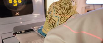
- the patient enters the office, protected from light and sound;
- an encephalograph “cap” consisting of special sensors is put on it;
- The sensor wires are connected to a device that records the bioelectric impulses of the brain.
Diagnostic stage
- the encephalograph transmits data to the monitor in the form of a graph;
- the power of electric fields and its distribution in different parts of the brain are recorded;
- Functional tests are carried out: the patient is asked to blink, look at flashes of light, breathe less often or deeper, listen to a sharp sound.
Final stage
- the electrodes are removed from the patient;
- print out the results.
Stages of implementation
The procedure for conducting an EEG is not particularly complicated and requires preliminary preparation. The patient has several requirements:
• the scalp should be clean. This will ensure better contact between the parting sensors and the patient's skin;
• for several days before the study, you should stop drinking alcohol or taking medications.
It is best to conduct research in the first half of the day. Small sensors fixed to an elastic mesh are attached to the scalp. Reference electrodes are fixed on the ears. Pulses are recorded for about 15 minutes.
After the EEG recording is processed and deciphered by the doctor who conducted the research, the result is given to the patient or transferred to his attending physician.
How long does the electroencephalogram procedure take?
A regular encephalogram (routine EEG or diagnosis of a paroxysmal state) takes from 20 to 30 minutes.
During the examination, a number of tests are carried out:
- rhythmic photostimulation;
- hyperventilation;
- load in the form of slow blinking.
If it is necessary to evaluate certain brain functions, the specialist adds additional tests, which he informs the patient about in advance. Such tests include:
- clenching your fingers into a fist;
- being in the dark;
- sleep deprivation for a certain period;
- night sleep monitoring.
Types of seizures and aura in epilepsy.
The international classification of epileptic seizures describes more than 20 types of seizures. In a simplified form, the classification looks like this:
- Partial (local) attacks occurring without disturbances of consciousness.
During these attacks, convulsive twitching involves some part of the body, can move from arm to leg or to the face and vice versa, but does not spread to the entire body. Sometimes attacks occur without convulsions. The manifestations of such focal attacks are varied: facial redness, drooling, flashes before the eyes, ringing in the ears. There is a special type of epilepsy - Jacksonian, in which seizures first seize one part of the body, then move to another in a certain sequence.
- Partial seizures occurring with disturbances of consciousness.
During these attacks, automatisms are common: a person unfastens buttons, begins to chew something, repeats the same phrase “what is it,” “I feel bad,” laughs or grimaces.
- Partial seizures with generalization.
These attacks begin as described above, then progress to a convulsive attack that involves the entire body.
- Generalized seizures.
These include non-convulsive and convulsive seizures.
Absence seizures are a type of non-convulsive seizure in which a person’s consciousness turns off for a short period of time. They may also be accompanied by automatisms and vegetative symptoms (redness of the face, drooling).
A generalized tonic-clonic seizure appears as a full-body seizure with loss of consciousness and amnesia (the person, upon regaining consciousness, does not remember the seizure itself or the events immediately preceding the seizure).
- In addition, there are atypical seizures that cannot be classified into any of the groups.
Sometimes an epileptic attack is preceded by an aura - a sensory sensation in the form of sound, color, smell, unpleasant taste, dizziness, mood changes, and abdominal discomfort.
The aura can manifest itself independently, without an attack. The type of aura is determined by the location of the epileptic focus in the cerebral cortex.
Interpretation of survey results
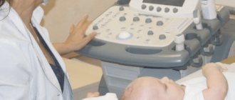
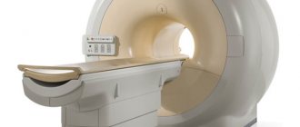
Even a qualified specialist is not always able to accurately name the cause of the patient’s poor health and immediately make a diagnosis. The doctor may be alerted to focal changes on the EEG. In this case, magnetic resonance imaging will be required to exclude a tumor or cyst.
When you contact a medical doctor, you will be able to undergo all examinations as quickly as possible, without delaying your visit to specialists.
If you need to do an EEG of the brain and you want to do it using modern expert-class equipment and in the shortest possible time, call the phone number listed on the website or leave a request in the feedback form.
Medical staff will answer your questions and conduct all necessary examinations within one business day. Let's take care of your health together!
Electroencephalography (EEG) is a method for recording spontaneous bioelectrical activity that occurs on the dendrites and bodies of pyramidal neurons of the cerebral cortex as a result of the summation of synchronous oscillations of inhibitory and excitatory postsynaptic potentials. This is the oldest method for assessing the functional state of the brain, first performed in humans in 1924 [1]. Before the widespread introduction of neuroimaging, EEG served to localize brain pathology, but the low spatial resolution, not exceeding 6 cm2, and the bends of the cerebral cortex, in which the electric field vector is parallel to the surface of the skull, make this indication unclaimed. Currently, the clinical use of the method is limited to the diagnosis of functional brain disorders [2–6].
EEG monitoring of patients with severe brain damage is the most dynamically developing area of clinical neurophysiology [7]. The high incidence of nonconvulsive status epilepticus (NSES) and ongoing differential diagnosis of motor activity in such patients make this type of continuous monitoring widely in demand in intensive care units (ICU) [8–10].
The procedure for analyzing EEG in patients with brain damage accompanied by depression of the level of wakefulness
EEG analysis is based on the assessment of the frequency and amplitude of oscillations of curves reflecting the bioelectrical activity of the brain, as well as verification of stereotypical graph elements. Similar to EEG data in epilepsy, in patients with primary and secondary brain damage, the recording protocol includes a description of background bioelectrical activity, epileptiform graphelements and patterns of rhythmic or periodic discharges. Each of them can have independent diagnostic and prognostic significance (Fig. 1).
Rice. 1. Scheme of the EEG protocol in a patient with depressed level of wakefulness.
Pathological changes in background bioelectrical activity in patients with suppressed levels of wakefulness due to brain damage are characterized by a diffuse, hemispheric or limited to several leads slowing of the background rhythm, a progressive decrease in the amplitude of oscillations, until their complete disappearance. A feature of EEG analysis in such patients is the frequent detection of a “gap rhythm” that occurs in the projection of a defect in the skull bones, and the need to assess reactivity in response to external stimuli. The lack of reactivity indicates an unfavorable prognosis for the restoration of consciousness [11, 12] (Fig. 2).
Rice. 2. EEG, abbreviated bipolar montage. Severe traumatic brain injury, depression of the level of wakefulness to moderate coma. Changes in the frequency and amplitude of the background rhythm in response to the touch of a patient’s relative (the moment of touch is marked with a vertical dotted line).
The sequence of background EEG signs and options for their description in patients with depression of wakefulness are presented in Table. 1.
Table 1. Scheme for describing the conclusion about the state of the background bioelectrical activity of a patient with depression of wakefulness. The description of single epileptiform discharges (ED), which occur in patients due to primary or secondary brain damage, do not differ from classical definitions, and all their types have the same diagnostic significance [ 13].
To assess the dynamics of the transition of sporadic ER into an epileptic seizure, an important factor is the gradation of the frequency of their occurrence. When drawing up an EEG conclusion, the following options for a textual description of ER are used: for sporadic ER, occurring more often than 10 s (one graph element on a standard EEG screen) - multiple; less than once every 10 seconds, and more often than once every 1 minute - frequent; less than 1 time per 1 minute, and more often than 1 time per 1 hour - episodic; less than once every 1 hour - rare.
In cases where the ER frequency exceeds 3.5 Hz, the overall picture of the EEG is considered as an electrographic epileptic seizure, and if it is not accompanied by pronounced motor manifestations, it is called non-convulsive.
Monomorphic EEG and ER graphelements, creating stereotypical patterns in the frequency range from 0.5 to 3.5 Hz, are found in more than 50% of patients in a coma. They pose the greatest challenge to clinical interpretation [14]. According to the terminology proposed by the American Clinical Neurophysiology Society (ACNS), they are called “rhythmic and periodic patterns” (RPP) [15]. In order for a combination of identical EEG graph elements to be recognized as an eating disorder, they must be recorded 6 times in a row or more.
To classify RPP, a special scheme is used, which includes a description of the localization and morphology of graph elements, as well as a number of additional features called modifiers (Fig. 3)
Rice. 3. Scheme for describing rhythmic and periodic patterns [14]. [15]. Its implementation made it possible to standardize the description of EEG, increasing the level of inter-expert agreement, and to conduct a number of cooperative studies that revealed the clinical significance of individual patterns [16, 17].
Methodology for describing rhythmic and periodic patterns
The terminology of eating disorders was proposed for discussion in 2005, and the final version was published in 2012 [15, 18]. It is included in the standard electronic system for generating EEG conclusions SCORE and translated into Russian [19]. To create databases, especially when conducting cooperative studies, it is necessary to clarify all potentially possible parameters, but in clinical practice only both basic terms should be indicated, and their modifiers should be used based on the principle of reasonable sufficiency.
Basic terms for describing RPP
Key term #1: Localization.
There are 4 possible options for its description:
1. Generalized (G)
. This is any bilateral, bisynchronous and necessarily symmetrical pattern (Fig. 4).
Rice.
4. EEG, reduced reference montage. The GPRs are outlined with a black rectangle. In case of amplitude dominance over one of the areas, the description is supplemented with one of the options: a) with frontal dominance - the amplitude of discharges in the frontal leads exceeds the occipital leads by 50% or more;
b) with occipital dominance - the opposite variant to the frontal one;
c) with central dominance - the addition is used when there is amplitude dominance in the parasagittal leads.
2. Lateralized (L).
These are unilateral or asymmetrical but synchronous bilateral patterns, including focal, regional and hemispheric (Fig. 5).
Rice.
5. EEG, abbreviated bipolar montage. Lateralized unilateral periodic discharges, spike-like (LPR + C). 3. Bilaterally independent (Bi). In this case, discharges occur over both hemispheres, but independently of each other.
4. Multifocal (Mf). The graphoelements of the pattern are recorded independently over different lobes of the brain.
The last two options require a description of the symmetry of the amplitude of the discharges, indicating the most involved lobe of the brain in each hemisphere.
Basic term No. 2. Morphology.
Described by one of three concepts:
— Periodic discharges (PR).
Periodic are any homologous graph elements that occur at close to the same interval, that is, an interval whose length varies within 50% in more than ½ cycles of the pattern. A discharge is a graph element with a duration shorter than 0.5 s and consisting of no more than 3 phases (Fig. 4). It should be distinguished from a flash, in which the curve crosses the isoline more than 3 times and the duration is from 0.5 to 4 s.
— Rhythmic delta activity (RDA).
Rhythmic is an activity consisting of repeating graphoelements with relatively the same morphology (monomorphic) without intervals between them. The oscillation frequency of the delta range does not exceed 4 Hz. A pattern meets the rhythmicity criteria if the duration of each cycle varies within 50% in the majority (>50%) of pairs of cycles.
— Spike-wave (SW).
A pattern consisting of a series of complex graph elements combining a spike (polyspike) or sharp wave followed by a slow wave that occurs constantly and at the same interval. The pattern is characterized by the absence of an interval between complexes, otherwise it is called “PR with spike morphology.”
Modifiers complement the basic terms of eating disorder and have different clinical meanings [20].
Modifiers of basic RPP terms
In the description of the RPP, modifiers are distinguished that clarify the morphology of graph elements, called “ modifiers plus”
"(Fig. 3) and additional terms characterizing the properties of the pattern (Table 2)
Table 2. Main RPP modifiers and their possible meanings [15].
1. Morphology modifiers
"+". They indicate a high probability of ictal pattern. Applicable only to describe PR or RDA [15].
Modifiers “+” are of the following types:
A. + Fast activity (B)
. Superposition of P.R. or RDA with oscillations exceeding a frequency of 4 Hz.
b. +Rhythmic (quasi-rhythmic) delta activity (R) - used only with the PR pattern.
V. + Spike, sharp or sharply defined wave (C)
- used with the appropriate form of RDA graphic elements.
d. Combinations of modifiers
—
+
Fast rhythmic (BR) for PR and
+
Fast spiky (FR) for RDA.
Pattern modifiers serve to clarify its ictal nature and assess the likelihood of transition from eating disorder to BSES. In order to increase Cohen's kappa, which reflects the degree of inter-rater agreement (IEA), unambiguous definitions were adopted to describe each of the modifiers. They are described in detail in Russian and English [15, 20].
To make a decision about starting intensive therapy, the frequency of graph elements in the pattern and its dynamics are most important.
Pattern dynamics
. One of the most important modifiers that must be used if the development of BSES is suspected.
1. Pattern with evolution
- at least two consecutive changes
in the frequency
,
morphology
or
localization
of its graph elements. The dynamics of these signs must satisfy the following criteria.
A . Frequency
- at least 2 consecutive identically directed frequency changes of 0.5 Hz or more. For example, gradual acceleration 2.5→3.0→3.5 Hz or deceleration 2.0←1.5←1.0 Hz of pattern graphic elements.
b . Morphology -
the sequential appearance of two RPP graphelements, each of which has a morphology different from the previous one.
V. Localization -
gradual spread of RPP to more than 2 adjacent electrodes installed according to the 10-20% scheme.
2. Pattern with fluctuation
— a dynamic state, including 3 or more changes in frequency, morphology or localization that do not meet the criteria of evolution, while the duration of static sections of the pattern should not exceed 1 minute. Thus, a pattern of RDA of 1.5 Hz for 30 s, passing into RDA of 2 Hz for 50 s with a return of frequency to 1.5 Hz for 30 s, is regarded as fluctuating, and PR in leads C3 and T3 for 40 s, then expanding presence on F3 for 3 min with reverse development on C3 and T3 for 50 s - no.
An isolated change in the amplitude of the RPP should be regarded as evolution or fluctuation.
3. Static pattern
- absence of any changes during the entire period of its registration.
The sequence of description and variant values of the remaining modifiers are presented in table. 2.
To create a database of studies and a detailed description of the EEG, it may be necessary to describe other, less significant features. They are called "minor modifiers". These include three-phase morphology, dynamics of occurrence and the presence of a fronto-occipital gradient of RPP.
A comparison of the new classification with the considered classical definitions of H. Lüders [22] is presented in table. 3 [21].
Table 3. Comparison of the most common eating disorders classified according to the ACNS scheme and their description according to Lüders
Standardized terminology for describing eating disorder made it possible to increase the MES to 0.81 and create a scientific community for the study of EEG monitoring data in ICU patients (Critical Care EEG Monitoring Research Consortium - CCEMRC) to conduct cooperative research. As a result, individual EEG patterns were identified that were specific to the prognosis of an unfavorable outcome of the disease, indicating BSES or the risk of its development.
Clinical significance of eating disorder
The pathophysiological processes leading to the appearance of eating disorder in patients with brain damage are not completely clear. It is assumed that they are caused by the development of primary metabolic disorders or disconnection of connections between neurons of the contralateral cortex, resulting from direct traumatic effects or hypoxia. Registration of eating disorder accompanies neuronal damage and worsens the prognosis of the course of brain damage [23, 24]. There are two views on their pathophysiology. According to the first, RPP are graph elements that only indicate diffuse damage to the cerebral cortex. The second considers them as a sign of an active pathological process, similar to an epileptic seizure, which causes a local increase in metabolism and leads to secondary damage to neurons [25].
The processes of epileptogenesis, which develop as a result of damage to the cerebral cortex and hemorrhage, also lead to the appearance of eating disorders, which may have an epileptiform morphology of graphoelements and developmental dynamics [26].
The result of the analysis of a number of multicenter cooperative studies was the identification of a number of eating disorders and their special characteristics specific to BSES, a high risk of developing an epileptic seizure and predicting the outcomes of secondary brain damage resulting from prolonged anoxia.
Status epilepticus is an epileptic attack without clinical signs of its cessation, with a duration exceeding most attacks of this type, or attacks that are repeated one after another, but without complete restoration of the background state of the nervous system between them. For each type of attack, the concept of “abnormal duration time” is different; it is conventionally called time t1. Continued status epilepticus leads to irreversible damage to neurons and destruction of interneuronal connections. The period after which they arise is called time t2 [27]. Indicators t1 and t2 are determined experimentally; they differ for different types of attacks. For example, in tonic-clonic status epilepticus, the times t1 and t2 are 5 and 30 minutes, respectively. The same time parameters are accepted for BSES, which developed in patients with severe brain damage [28].
Diagnosis of convulsive status epilepticus is not difficult due to the clinical picture of the convulsive syndrome. The development of BSES occurs subclinically and is accompanied only by a change in consciousness or depression of wakefulness. Status epilepticus is recorded in 5-48% of patients in a coma of any etiology [29]. In work [29], conducted at the Department of Emergency Neurosurgery of the Research Institute for Emergency Medicine named after. N.V. Sklifosovsky, it was found that in the acute period of severe traumatic brain injury, BSES develops in 28% of victims.
In patients with diseases leading to depression of wakefulness to the point of coma, or during therapeutic sedation, clinical assessment of changes in consciousness is not possible. Therefore, the diagnosis of BSES is based on identifying EEG signs of an epileptic seizure and classification of eating disorders, followed by analysis of the relationship of these changes with the patient’s clinical condition.
In 2013, S. Beniczky et al. [26] presented clinical and electrographic signs of BSES. Thus, the diagnostic criteria for BSES in patients who do not suffer from epilepsy are: 1) PR frequency more than 2.5 Hz; 2) PR frequency less than 2.5 Hz or RDA pattern and one of the following signs: a) electrographic (restoration of background rhythm and its reactivity) and clinical improvement with the administration of antiepileptic drugs (AEDs); b) minor clinical ictal signs during recording of the electrographic pattern; or; c) evolution of PR or RDA. They were subsequently validated in a cooperative study using standardized eating disorder terminology [30].
The diagnosis of BSES is considered established with clinical suspicion of this condition and identified EEG changes that exactly meet the criteria presented by S. Beniczky [26]. In the case of EEG improvement in response to the introduction of AEDs or a fluctuating pattern of PR and RDA, they speak of probable BSES.
Diagnosis and control of treatment of BSES are possible only with the help of EEG. The EEG pattern of status epilepticus does not differ from the changes that are recorded during a single epileptic seizure (Fig. 6).
Rice. 6. EEG, abbreviated reference montage. Lateralized bilaterally asymmetrical LRDA +B, evolving pattern (boxed). The typical evolution of the frequency of RDA without clinical manifestations in the form of seizures indicates BSES.
Prediction of convulsive and non-convulsive seizure/status based on dynamic analysis of eating disorder
Bedside EEG monitoring makes it possible to detect not only BSES, but also other eating disorders that are specific to a high risk of epileptic seizures [31, 32]. Patterns differ in morphology, frequency and dynamics of distribution in space and time, and their occurrence and transition to an electrographic or clinical epileptic seizure are considered as a single process called the “interictal-ictal continuum” [33, 34]. All rhythmic patterns (SV and RDA) with a frequency of graph elements exceeding 3 Hz, and periodic patterns accompanied by minor rhythmic movements of the muscles of the face or limbs, are considered as an unambiguous sign of an electrographic epileptic seizure [26]. The need to initiate treatment for eating disorder with a lower frequency of graph elements and the ratio of risk to possible positive outcome remains controversial. Delayed initiation of treatment for BSES worsens functional outcomes, but the unjustified use of sedating medications used to treat refractory forms of BSES is accompanied by prolonged artificial ventilation, which increases the risk of infectious complications [33].
A. Bauerschmidt et al. [31] proposed an approach to determining indications for eating disorder therapy in patients with depressed levels of wakefulness, based on an analysis of the frequency and morphology of pattern graphoelements, as well as their quantitative assessment and response to intravenous administration of benzodiazepines:
— Frequency of graph elements in the RPP
. The lower limit from which therapy should begin is considered to be 2 Hz. Patterns with a frequency of 0.5 to 2 Hz require treatment only if there are rhythmic, stereotypical movements that coincide with the pattern on the EEG.
— Morphology of graph elements
. All lateralized RPP (LPR, LRDA, LSV) indicate the risk of transition to an epileptic seizure, however, their analysis using the “+” modifier allows for a more accurate prediction of its development. The highest probability of an epileptic seizure was noted when registering the DM with the modifier +B, +S and +BS and the intervals between graph elements less than 1 s [33].
— Quantitative assessment of RPP with calculation of a special index.
Two quantitative methods have been proposed to assess the risk of developing an epileptic seizure in patients with severe brain damage. The first, called the 2HELP2B scale by the authors, consists of assigning a certain number of points to 6 signs that describe an eating disorder. According to the 2HELP2B scale, which assesses the risk of developing an attack based on EEG data, short-term ictal rhythmic discharges correspond to 2 points; Decision maker or LRDA or BiPR - 2; epileptic seizure before the start of EEG recording - 1; sporadic ER - 1; frequency of graph elements in the RPP above 2 Hz - 1; the presence of any modifier “+” – 1 point. The risk of an attack for 0 points is 5%, for 1 point - 12%, 2 points - 27%, 3 points - 50%, 4 points - 73%, 5 points - 88%, 6 and 7 points - 95% [35] . The second method is to use the GP scale, which includes a history of epilepsy (1 point), focal changes on the EEG (2 points), and the absence of triphasic morphology of the PD (3 points) [36]. The risk of an attack for patients with HPR who did not score points on the scale is 13%, 5-6 points - 94%.
— Reaction to benzodiazepines
. Electrographic and clinical improvement with intravenous administration of AEDs is one of the diagnostic signs of BSES. The same test can be used to decide on the initiation of intensive prophylaxis of status epilepticus when an eating disorder of unclear diagnostic significance is detected [37]. Preference is given to fast-acting benzodiazepines, such as diazepam or midazolam, and the dose of the drug should not exceed the therapeutic dose. The disappearance of eating disorder, restoration of background rhythms and the appearance of reactivity on the EEG indicate the need to begin preventive therapy with anticonvulsants.
Differential diagnosis of RPP ictality can be supplemented by assessment of the results obtained using functional neuroimaging methods, such as diffusion-weighted magnetic resonance imaging (DW-MRI), fluorodeoxyglucose positron emission tomography (FDG-PET), single-photon emission computed tomography (SPECT) ) or according to tissue microdialysis data [23, 38, 39].
In Fig. 7 presented
Rice. 7. Decision-making scheme for eating disorder therapy with a frequency less than 3 Hz. BDZ - benzodiazepines. scheme for the analysis of eating disorders, the use of additional diagnostic methods and drug tests to identify eating disorder of ictal nature, requiring intensive therapy, proposed by A. Bauerschmidt et al. [31].
Prediction of outcomes of diseases accompanied by depression of wakefulness to coma, according to analysis of background EEG and RPP
Numerous studies have established the high prognostic value of EEG in patients with secondary brain damage resulting from prolonged hypoxia after restoration of cardiac activity [40].
EEG signs of an unfavorable course of postanoxic encephalopathy are a violation of the constancy of bioelectrical activity to the level of the “flash-suppression” pattern, a decrease in the background amplitude below 10 µV/mm, the disappearance of EEG reactivity and variability. Among RPP, GPR with a low frequency of discharges in the pattern against the background of completely suppressed background activity is considered “malignant”. The sensitivity of these signs for death or transition to a permanent vegetative state is 50%, specificity is 100% [41-43].
Prediction of survival and recovery of consciousness in patients with primary brain damage is also based on analysis of background bioelectrical activity of the brain and classification of eating disorders, but their information content has not been sufficiently studied.
Preservation of reactivity of EEG patterns in victims with severe traumatic brain injury is a positive prognostic sign with a specificity of 89%, but its absence does not indicate an unfavorable prognosis [12, 44].
Among RPP, only GPR is considered a sign of an unfavorable course of primary brain damage, but its sensitivity and specificity have not been established [45].
The decision to conduct a standard, extended or computer EEG and change treatment tactics based on its results should be made only taking into account the clinical picture and neuroimaging data. Indications for EEG in patients with severe brain damage, adopted by the international consensus conference on multimodal neuromonitoring in the ICU, are presented in Table. 4 [46,
Table 4. Recommendations for the use of EEG in a complex of multimodal monitoring in conditions of neurocritical care 47].
For correct clinical interpretation of EEG results and timely initiation of intensive anticonvulsant therapy, standardized terminology for describing eating disorder should be used. The minimum necessary is a description of the localization and morphology of the graphoelements of the pattern and modifiers that reflect the ictal nature of the RPP - the frequency of the discharge in the pattern, and its dynamics.
The authors declare no conflict of interest.