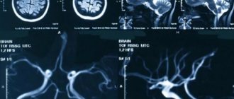
Ultrasound of cerebral vessels
Dopplerography of cerebral vessels is prescribed for suspected neurological diseases. The following symptoms may indicate this:
- Panic attacks.
- Difficulty speaking.
- Difficulties in perceiving information.
- Impaired coordination of movements.
- Decreased visual, auditory, kinesthetic sensations.
Based on the results of the ultrasound, the doctor can determine what changes in the structure of the blood vessels could cause these symptoms and prescribe treatment, if necessary.
The procedure is simple and safe and has no age restrictions. It can be carried out at home using a portable scanner.
Who is the procedure indicated for?
Ultrasound examination is prescribed for those patients who express complaints:
- For cephalgia.
- Dizziness.
- A sharp decrease in vision.
- Fainting conditions.
- Memory loss.
- Loss of coordination.
- The appearance of dark circles and spots before the eyes.
- Insomnia.
- Decreased facial skin sensitivity.
- Excess body weight.
- A sharp decrease in vision.
- General weakness and fatigue.
- Numbness of the upper extremities.
- Hand tremors.
- Increased blood pressure caused by kidney disease.
In addition, people who have suffered head injuries, strokes and heart attacks are referred for brain ultrasound.
This diagnostic method is used in the presence of high levels of cholesterol in the blood, various types of arrhythmia, cervical osteochondrosis, stenosis, atherosclerosis, arterial hypertension, and coronary artery disease.
In addition, ultrasound examination is prescribed after vascular operations, as well as to monitor the therapy.
The essence of Doppler ultrasound
Based on the name of the procedure, it is performed using ultrasonic waves. Their work is based on the Doppler effect. The procedure differs from a standard ultrasound: a vessel with blood passing through it is displayed on the monitor screen. Looking at the resulting image, the specialist assesses the state of blood flow and vascular patency, which affects the quality of blood supply to the brain.
Dopplerography of cerebral vessels makes it possible to study the work of the veins, which “supply” blood to the brain, and the arteries, which are responsible for the outflow of waste products from it. The vessels being examined are located in the cervical and cranial regions. The type of ultrasound will depend on their location:
- Dopplerography of extracranial vessels located on the surface of the neck (jugular veins, carotid, vertebral and subclavian arteries). Any disturbances in their structure affect the state of the brain, which is why these vessels are called “maternal” vessels.
- Dopplerography of transcranial, or “daughter” vessels. The doctor installs an ultrasound sensor in the thinnest parts of the skull and evaluates the functioning of the veins and arteries that supply the brain.
Prices for services
- Diagnostics
Note! Prices are for adult patients. Please see the “Pediatrics” section for the cost of children’s appointments.
| Ultrasound of the abdominal organs (liver, gall bladder, pancreas, spleen) | 1700 |
| Ultrasound of the abdominal organs (liver, gall bladder, pancreas, spleen) + kidneys | 2500 |
| Ultrasound of the gallbladder with functional tests | 1940 |
| Ultrasound of the liver and gallbladder | 1500 |
| Kidney ultrasound | 1500 |
| Ultrasound of the adrenal glands | 800 |
| Ultrasound of the bladder | 970 |
| Ultrasound of the scrotum | 1600 |
| Ultrasound of the bladder and prostate | 1340 |
| Ultrasound of the bladder and prostate with determination of residual urine | 1820 |
| Ultrasound of the prostate gland transrectally | 2100 |
| Ultrasound of the pelvic organs with an abdominal sensor (gynecology, urology) | 1600 |
| Ultrasound of the pelvic organs with a vaginal sensor (gynecology) | 2000 |
| Folliculometry (ultrasound of follicles) | 1100 |
| Ultrasound of the thyroid gland with determination of blood flow | 1600 |
| Ultrasound of the mammary glands with determination of blood flow | 2000 |
| Ultrasound of soft tissues | 1400 |
| Ultrasound of lymph nodes (1 area) | 1500 |
| Ultrasound of the heart in color Doppler mode (echocardiography) | 3500 |
| Ultrasound (duplex) of veins of the extremities | 2600 |
| Ultrasound of the brain (neurosonography in children) | 1700 |
| Ultrasound of cerebral vessels (duplex scanning) | 2700 |
| Ultrasound of neck vessels (extracranial) | 2700 |
| Ultrasound of the knee joints (2 joints) | 2490 |
| Ultrasound of the hip joints (2 joints) | 2700 |
| Ultrasound of small (elbow, wrist, ankle) joints (1 joint) | 1460 |
| Fine-needle aspiration biopsy (FNA) of a thyroid cyst/nodule under ultrasound control (1 cyst), without the cost of histology | 3340 |
| Ultrasound of the sinuses (maximum) | 1200 |
| Ultrasound of the eyeball and orbital structures (1 eye) | 1460 |
| Ultrasound measurement of the anteroposterior segment of one eyeball | 1000 |
| Ultrasound of the parathyroid glands | 1200 |
| Ultrasound of the spleen | 1200 |
| Duplex scanning of the renal arteries* | 2700 |
| Duplex scanning of neck and brain vessels* | 4400 |
| Hysterosalpingography (HSG)* (without the cost of manipulation) | 2790 |
| Doppler ultrasound of the abdominal aorta* | 2200 |
| Doppler ultrasound of the renal arteries* | 2700 |
| Ultrasound of the stomach* | 2010 |
| Ultrasound of one organ* | 1200 |
| Ultrasound of pleural sinuses* | 1100 |
| Abdominal ultrasound + study of blood flow of the liver and abdominal aorta* | 2500 |
| Ultrasound of the lungs, bronchi and pleural cavities | 2500 |
In what cases is ultrasound ultrasound prescribed?
The main symptoms signaling a cerebrovascular accident are:
- Impaired coordination of movements.
- Deterioration of hearing and memory.
- Frequent attacks of headache.
- Impairment or partial loss of taste.
- Insomnia, difficulty falling asleep.
- Noise, whistling and ringing in the ears.
- Deterioration of visual function, manifested in loss of visual fields.
- Difficulty concentrating.
- Decreased sensitivity and motor activity of the arms and legs.
A doctor may prescribe a procedure in the following cases:
- The patient has diabetes mellitus.
- For arrhythmia, as this increases the risk of cardiac clot rupture and artery blockage.
- The patient has suffered a heart attack or stroke.
- Osteochondrosis of the cervical spine.
- Before planned heart surgery.
- If the patient is a smoker with many years of experience.
- An area with strong pulsation is visible on the neck (in this case, an ultrasound of the neck is prescribed).
Based on the above symptoms, the doctor can determine the location and name of the vessel in which the patency is impaired. Several pronounced symptoms indicate poor blood flow in a large vessel. In this case, extracranial Doppler ultrasound is prescribed. Violation of only one function will be the reason for conducting transcranial Doppler ultrasound.
Ultrasound of head and neck vessels
Ultrasound of the vessels of the neck and head is one of the most accessible and effective methods for diagnosing diseases such as atherosclerosis and thrombosis. The examination allows you to assess the condition of the arterial and venous branches and lymph nodes, measure the speed of blood flow and other parameters. Medical invites patients to undergo ultrasound diagnostics using new generation devices. An informative and safe examination will allow timely detection of vascular pathologies and initiation of treatment.
Indications for ultrasound examination of the vessels of the neck and head
An examination is prescribed if the patient notes the following symptoms:
- recurrent headaches, migraines;
- fainting conditions;
- dizziness not associated with hunger or fatigue;
- feeling of heaviness in the head in the morning after waking up;
- sleep disorders, insomnia;
- noise or congestion in the ears;
- sudden deterioration of vision;
- decreased concentration, memory problems;
- impaired coordination of movements;
- chronic fatigue, etc.
In addition, ultrasound examination of the vessels of the neck and head is carried out for such diagnoses and conditions as:
- traumatic brain injuries;
- stroke or heart attack;
- ischemic disease;
- hypertension;
- arrhythmia, angina pectoris;
- thrombosis of veins or arteries.
An ultrasound of the vessels of the neck and head will also be required if changes appear in blood tests, for example, if cholesterol levels increase. An annual examination is recommended for patients over 45 years of age. People who smoke a lot, lead a sedentary lifestyle, or work under constant stress are also at risk.
Contraindications
The procedure is quick and painless. Ultrasonic waves do not have a negative effect on the body and do not cause side effects. Thanks to these qualities, examination can be prescribed even to pregnant women and newborn children. The only limitation for diagnostics may be severe damage to the skin in the area of contact with the ultrasound equipment sensor.
Preparation for ultrasound of the vessels of the head and neck
Before undergoing the examination, the patient is recommended to consult with the attending physician. The specialist will most likely stop taking certain medications that may affect the functioning of blood vessels and distort the picture of the disease.
If the patient is involved in professional sports or heavy physical work, then on the eve of the examination the load should be minimized.
Preparing for ultrasound diagnostics also includes giving up coffee, strong tea, energy drinks, alcohol and smoking. It is not recommended to eat before the test.
How is ultrasound of the vessels of the brain and neck performed?
The main type of instrumental diagnosis of diseases of the cervical spine is a complex of duplex and triplex scanning. It gives an idea of the characteristics of blood flow in the vessels of the neck and head (including intracranial). During diagnostics, a complete picture of soft tissues is displayed on the equipment monitor, against which veins and arteries are clearly visible. The method allows you to record the speed of blood flow in a certain area. This parameter is coded in different colors: shades of red indicate the movement of blood towards the sensor, shades of blue indicate the movement of blood away from the sensor. The study makes it possible to identify such disorders as inhibition of blood flow due to atherosclerotic plaques, thinning of vessel walls, expansion or narrowing of the lumen, inflammatory processes, vascular damage due to diabetes or hypertension, congenital anatomical abnormalities in the structure of veins and arteries, and others.
The procedure takes from 30 to 60 minutes depending on the type of ultrasound and takes place in several stages:
1
The patient takes off his outer clothing, frees his neck and collarbones from jewelry, and then lies down on the couch.
2
The doctor sets up the equipment and treats the sensor with a colorless hypoallergenic gel, which can additionally be applied to the skin in the area of study. Acoustic gel improves the conductivity of the ultrasound signal and the gliding of the scanner.
At the end of the examination, the results are deciphered and given to the patient in writing.
What does an ultrasound scan show?
During the examination, the specialist examines the main arteries that provide nutrition to the brain: common, external and internal carotid, as well as middle, anterior, posterior vertebral and basilar.
The ultrasound protocol contains a detailed description of the anatomical features of the vessels of the patient’s neck and head, data on the movement of blood flow, obstruction or deformation of veins and arteries. The examination allows you to detect the following abnormalities and pathologies:
- Atherosclerotic plaques. At the initial stage of blockage formation, a thickening of the vessel wall of up to 1.5 mm is noted. A reading greater than this value may indicate the presence of plaque.
- Thrombosis. When the disease occurs, the protein fibrin and blood cells, platelets, accumulate in certain areas of the vessels. A thrombus can partially or completely block the lumen of a vein or artery, which leads to impaired circulation, hypoxia and other dangerous consequences.
- Cerebrovascular accident. Pathology can occur as a result of thrombosis, narrowing of the diameter of arteries and veins, changes in blood flow speed, development of osteochondrosis, etc. Identifying hypoxia at an early stage will prevent the occurrence of concomitant diseases, such as stroke.
Many vascular pathologies are irreversible, so patients at risk are required to undergo an annual ultrasound of the vessels of the head and neck - you can do this at our medical center in Moscow.
Advantages of the Miracle Doctor clinic
Experienced doctors
Ultrasound diagnostics of blood vessels is carried out by specialists with extensive practical experience. The total work experience of some doctors exceeds 30 years.
Modern equipment
The clinic has new generation ultrasound machines. The equipment allows you to obtain detailed images of blood vessels without interference.
All types of ultrasound at low prices
By contacting us, you can undergo Doppler ultrasound, duplex and triplex scanning of blood vessels. Ultrasound diagnostics are performed by appointment. To get tested, make an appointment by phone. A description of the examination results is performed in front of the patient.
VIP service
You can undergo a vascular examination any day of the week without a queue. If necessary, we will schedule you for a consultation with a phlebologist or neurologist.
To find out the cost of ultrasound of the vessels of the head and neck, make an appointment and ask other questions you are interested in, call our clinic in Moscow or fill out the feedback form.
Rules for preparing for research
On the day of the procedure, you must refrain from:
- Taking antispasmodics such as “Riabal”, “No-shpa”, “Drotaverine”, “Papaverine”, “Baralgin”, “Cinnarizine”.
- Smoking.
- Drinking black tea and drinks containing caffeine.
- Staying in unventilated, stuffy rooms with large crowds of people, which can have a detrimental effect on vascular tone.
If the patient is undergoing treatment with cardiovascular medications, then the advisability of discontinuing them before the procedure must be agreed with a neurologist. In any case, the sonologist must be informed about the treatment being performed before the ultrasound.
Stages of extracranial Dopplerography:
1. In the office, remove all jewelry from your neck and head.
2. Remove clothing from those parts of the body through which the sensor will be passed: neck, collarbones, shoulder blades.
3. Lie on the couch with your head facing the monitor. The doctor will apply acoustic gel to open areas of the body and begin to move the device over them, placing it in areas where medium and large arteries and veins are located. To obtain more accurate data on the state of vascular tone, the patient will need to hold his breath at the doctor’s request, take special medications and change body position.
Doppler ultrasound of vessels located in the cranial area has a different specificity. Acoustic gel is applied to the temples, the back of the head and the area above the eyes. The device is installed there because in these places the bones of the skull are the thinnest and are able to transmit ultrasonic waves.
By what principle is the received data decrypted?
Each vessel has its own parameters, on the basis of which calculations are made and the degree of their discrepancy with the norm is assessed.
Transcranial and extracranial Dopplerography of arteries compares digital data in each vessel segment:
- Artery wall thickness.
- Vessel diameter.
- Phases of blood flow, its features.
- The degree of symmetry of blood flow through identical arteries located on different sides.
- Diastolic blood velocity.
- Maximum systolic blood flow velocity.
- Pulsation and resistive indices, systole and diastole ratio.
- The degree of pathological narrowing of blood vessels (stenosis), the condition of the artery beyond its limits.
Dopplerography of veins evaluates the following parameters:
- Condition of the venous wall of the vessel.
- The nature of blood movement through the studied vessels.
- Vein diameter
Data obtained at rest and after functional tests are compared with normal values. Any deviations are marked with a special abbreviation of letters and numbers. Based on them, the neurologist prescribes further studies or forms a course of treatment.
Where can I get Doppler ultrasound of cerebral vessels?
The study is carried out in municipal clinics and public hospitals, specialized clinics, and general medical centers. Depending on the location of the ultrasound, the cost of each type of Doppler ultrasound will be in the range of 500-6000 rubles. The procedure has many advantages, including painlessness and the absence of side effects. Within 20 minutes, a sonologist will be able to determine the presence of anomalies in venous or arterial inflow and assess the degree of development of danger to blood vessels.
However, the procedure will not help determine the cause of compression of the thin arteries, since the capabilities of the device do not allow them to be seen from the outside.


