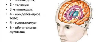
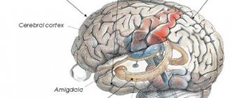
Structures associated with the limbic system are located in the inner part of the temporal lobe of the brain: gyrus hippocampalis, which goes rostrally into the uncus and includes the amygdala and hippocampus, are involved in the regulation of the functions of the autonomic nervous system and the affective sphere. These areas of the brain are also credited with being responsible for drive and motivation, memory, and learning.
The limbic system of the brain is known to be associated with the emotional sphere of a person; its damage in schizophrenia is quite acceptable, given the mood swings, manic states, depression and agitation observed in this disease.
It is possible that in schizophrenia, not all components of the limbic system are affected equally, but more subtle morphological changes may occur in the structure of the neurons included in this system. Some, but distinct, of these changes have been found in some cases in patients suffering from bipolar affective disorder.
The results of many studies in schizophrenia show a decrease in the density of neurons in the hypothalamus, amygdala-hippocampus complex and parahippocampal gyrus.
The dimensions of the parahippocampal gyrus and entorhinal cortex, according to K. Prasad et al. (2004), find a certain correlation with the severity of delusions and psychotic symptoms in schizophrenia. According to researchers, these brain structures play an important role in regulating processes related to memory.
A reduction in size and change in the shape of neurons was found in the hippocampus, hippocampal gyrus and entorhinal cortex (Bogerts B., 1993).
Hippocampus
In a study by C. McDonald et al. (2006) revealed a decrease in the volume of the right and left hippocampus, 2.47 ml and 2.50 ml, versus 2.54 ml and 2.61 ml in healthy individuals, which corresponds to approximately a decrease in the volume of this brain structure by approximately 6%.
Some authors note that a decrease in the volume of the hippocampus, as part of the limbic system, is noticeable after the first psychotic episode, however, according to other researchers, these changes are recorded before the manifestation of schizophrenia and progress after its onset. Note that in relatives of patients with schizophrenia, a decrease in the volume of the hippocampus and amygdala can also be detected.
In schizophrenia, the functioning of the amygdala is impaired; this fact is discovered when studying patients with schizophrenia using the method of evoked potentials (P300).
In the hippocampus of patients with schizophrenia, with a decrease in the volume of neurons and “sparseness” of their mutual arrangement, an increase in the number of pathologically altered myelinated axons with thinned myelin sheaths, swollen perioxal glial processes and wrinkled axons was found. The proportion of these fibers in the total number of myelinated axons and their numerical density in schizophrenia is 2 times greater than in the control group. At the same time, it is known that pathological and reparative changes in axons depend on the reaction of the microglial cells surrounding them (Kolomeets N.S., 2007).
Peculiar memory impairments, primarily working memory, in schizophrenia, affecting learning difficulties in persons suffering from this disease, may indicate the involvement of the hippocampus in the pathological process.
According to Shenton et al. (2001), the volumes of the medial temporal lobes, which usually include the hippocampus and amygdala, are significantly reduced in size. These changes were noted in 70% of patients with schizophrenia.
Limbic system (continued)
According to James, emotions are associated with the activity of the sensory neocortex, as a place of integration of body sensations reproduced in emotional states. The fact, or rather the concept that certain brain structures can form emotional states (emotional systems of the brain) was somewhat unexpected for neurologists and, in fact, was a kind of launching pad for such a discipline as biological psychiatry and “behavioral neurology” - branches of neurology related to how neurological disorders are associated with behavioral changes.
In 1937, Papez described a proposed cerebral mechanism of emotion. Key components of his now well-known circuit were the hippocampus, cingulate cortex, mammillary bodies, and anterior thalamus. For Papez, the cingulate cortex was the receptive area for experienced emotions, in the same way that the visual area of the brain was receptive for visual information. The hypothalamus was the basis for the expression (expression) of emotions, and to experience them, the cerebral cortex was necessary, the “flow of feelings” depended on the strength of the connections between the cortex and the hypothalamus. In conclusion, Papez summarized, in a now well-known quote, that “the hypothalamus, the anterior thalamic nuclei, the cingulate cortex, the hippocampus and their interconnections form a harmonic mechanism that can represent a functionally complex central emotion and also participate in emotional experience.
The key structures of the limbic system, originally outlined by MacLean, but developed in detail by other authors, were the amygdala and hippocampus - a kind of neural aggregates functioning in a complex with directly related structures such as the orbital part of the frontal cortex and the so-called ventral striatum. - part of the extrapyramidal system of the brain, which is related to the motor component of emotion (“motor expression or expression of emotion”). In humans, due to the migration of the temporal lobe to the posterior lower parts of the brain and the large size of the corpus callosum, the archicortex (evolutionarily old cortex) is, as it were, folded and closed in the medial part of the temporal lobe - in the hippocampus.
The cingulate gyrus surrounds the corpus callosum, forming a C-shaped group, connected in the posterior part to the parahippocampal gyrus and extensively in contact with neocortical structures. The frontal lobe of the brain includes a large number of demarcacinal parts (subregions), but the most active and well-known are the orbital, medial and dorsolateral regions of the frontal lobe. The orbitofrontal cortex is located superior to the cranial fossa and has intimate connections with the anterior insula, amygdala, ventral striatum, and sensory projection tracts. The ventromedial frontal cortex is part of the cingulate cortex. The insula is a relatively large limbic structure that, in contrast to most components of the limbic system, remains invisible from the medial surface of the brain and lies laterally and is contained (closed) below the flexures of the neocortex.
Many researchers are critical of the term "limbic system", and some anatomists today prefer to use the term "limbic lobe". after the original Broca designation, thereby destroying the concept of the “limbic system”. With the development of such modern techniques of neuroanatomy as the use of horseradish peroxidase (HRP), histofluorescence, autoradiography and the modernized method of silver staining, proposed in the early stages of the development of neuroanatomy, interest in the structural and functional features of the limbic lobe, especially cortical-subcortical relationships, has grown significantly. in particular, in terms of the development of ideas about the neuroanatomy of emotions and behavior.
Although the Broca term is largely topographical, the limbic lobe can be differentiated from other cortical structures by cell staining and vulnerability to certain neuropsychiatric disorders. In one of the latest definitions, in particular those proposed by Heimer et.al. (2007), the limbic lobe consists of the olfactory allocortex, the hypocampal allocortex, and transitional cortical areas that communicate with the larger isocortex. Transitional areas are quite numerous and form the majority of the limbic lobe and are separated in terms of “structure” by one or more boundaries (tracts) from the isocortex. The cortical fields of the limbic lobe form the cingulate and parahippocampal gyri, well connecting or connecting the caudal orbitofrontal, medial frontal, temporopolar, anteroventral part of the insula and the retrosplenial cortex, and also include the laterobasal-cortical complex of the amygdala.
An expanded original concept of the limbic system was proposed by Nauta, Domenesick (1982), who included the posterior orbito-frontal cortex and the temporal pole, which gives projections to the amygdala and hippocampus, as well as some subcortical structures interconnected by reciprocal pathways. They include the accumbens (nuclei accumbens) and midbrain structures such as the ventral tegmental area (VTA) and intepeduncular nuclei. The inclusion of these structures led to the formation of the concept of the midbrain limbic region of the mesolimbic system.
Nienwenhuys (1996) at one time coined the term “large limbic system,” which also included the pons and medullary neuronal group related to autonomic and somatomotor integration. He emphasized that this system consists of a mass of tonic and ultrafine fibers (fibres) and includes neuronal groups related to monoamine and peptide activity and contains places (foci) with the activity of which certain behavioral patterns can be associated.
It is important to understand that anatomically the limbic lobe is not an isocortical structure, does not have a six-layer structure, but consists of an allocortex and a belt of agranular or dislaminar (the 4th field is absent, poorly organized or atypical) intermediate cortex. In other words, possible definitions of the limbic cortex are related to those areas of the cortex that are not isocortical (six-layer).
Input pathways to the limbic system can be either interoceptive (visceral) or exteroceptive (transmitting information from the environment). Formation from different structures provide information about the internal environment of the body and include the modulating influence of neurotransmitter tracts, which originate from the midbrain and hindbrain and provide drive for behavior and modulation of emotional state through dopamine, serotonin and norepinephrine. Exteroceptive afferent connections from all sensory systems ultimately comprehensively integrate sensory information to the hippocampal cortex and amygdala.
Almond nuclei
In a series of brain autopsies of patients with schizophrenia, no significant increase in the volume of the amygdala nuclei was detected (Steven A. et al., 2002). However, data obtained using MRI currently refers to rather crude imaging methods, not to mention CT, and may be less reliable compared to autopsy.
In a comparative review by Wright et al (2000), who studied the size of 44 brain regions in schizophrenia, it was noted that the amygdala of the left and right hemispheres was reduced in volume by 10%, and this was noticeable to a greater extent than the reduction in volume of other brain regions. Loss of gray matter in the amygdala and changes in the shape of the amygdala have also been demonstrated through studies that looked at ventricular-brain index scores. At the same time, many authors note methodological difficulties in studying the amygdala of the limbic system of patients with schizophrenia.
Return to Contents
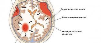
White matter
It is presented in the form of numerous fibers. They are divided into three groups:
- Projection. This category is represented by bundles of efferent and afferent fibers. Through them, there are connections between the projection centers and the basal, stem and spinal nuclei.
- Associative. These fibers provide connection to the cortical areas within the boundaries of one hemisphere. They are divided into short and long.
- Commissural. These elements connect the cortical zones of the opposite hemispheres. Commissural formations are considered: the corpus callosum, posterior and anterior commissure and commissure of the fornix.
Surface of the raincoat element
In each segment, this part of the brain is divided into lobes by deep grooves and fissures. Primary are referred to as permanent formations of the organ. They are formed at the embryonic stage (in the fifth month). The largest fissures include longitudinal (separates the segments) and transverse (separates the cerebellum from the occipital lobes).
Secondary and especially tertiary formations determine the individual relief of the segments (it can be seen in the photo). The human brain develops not only during the prenatal period. For example, secondary and tertiary grooves are formed up to 7-8 years after birth. The relief that the telencephalon has, the location of permanent formations and large convolutions are similar in most people. Each segment has six lobes: limbic, insular, temporal, occipital, parietal and frontal.
The telencephalon in this area includes the Rolandian (central) sulcus. With its help, the parietal and frontal lobes are separated. There is also a Sylvian (lateral) fissure on the surface. Through it, the parietal and frontal lobes are separated from the temporal lobe. A conventional line acts as the anterior-inferior border of the occipital region.
Neocortex
The neocortex is a part of the brain found in higher mammals. The rudiments of the neocortex are also observed in lower animals that suck milk, but they do not reach high development. In humans, the isocortex is the lion's part of the general cerebral cortex, having an average thickness of 4 millimeters. The area of the neocortex reaches 220 thousand square meters. mm.
History of origin
At the moment, the neocortex is the highest stage of human evolution. Scientists were able to study the first manifestations of the neobark in representatives of reptiles. The last animals in the chain of development without a new cortex were birds. And only humans have a developed neural system.
Evolution is a complex and long process. Every species of creature goes through a harsh evolutionary process. If an animal species was unable to adapt to a changing external environment, the species lost its existence. Why was man able to adapt and survive to this day?
Being in favorable living conditions (warm climate and protein foods), human descendants (before the Neanderthals) had no choice but to eat and reproduce (thanks to the developed limbic system). Because of this, the mass of the brain, by the standards of the duration of evolution, gained a critical mass in a short period of time (several million years). By the way, the brain mass in those days was 20% greater than that of a modern person.
However, all good things come to an end sooner or later. With a change in climate, descendants needed to change their place of residence, and with it, start looking for food. Having a huge brain, descendants began to use it to find food, and then for social involvement, because. It turned out that by uniting into groups according to certain behavioral criteria, it was easier to survive. For example, in a group where everyone shared food with other members of the group, there was a greater chance of survival (Someone was good at picking berries, someone was good at hunting, etc.).
From that moment on a separate evolution for the brain , separate from the evolution of the whole body. Since those times, a person’s appearance has not changed much, but the composition of the brain is radically different.
What does it consist of?
The neocortex is a collection of nerve cells that form complex gray matter. Anatomically, there are 4 types of cortex, depending on its location - parietal, occipital, frontal, temporal. Histologically, the cortex consists of six balls of cells:
- Molecular ball;
- external granular;
- pyramidal neurons;
- internal granular;
- ganglion layer;
- multiform cells.
What functions does it perform?
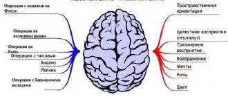
The human neocortex is classified into three functional areas:
- Sensory . This zone is responsible for higher processing of received stimuli from the external environment. So, ice becomes cold when information about the temperature arrives in the parietal region - on the other hand, there is no cold on the finger, but only an electrical impulse.
- Association zone . This area of the cortex is responsible for information communication between the motor cortex and the sensitive one.
- Motor zone . All conscious movements are formed in this part of the brain. In addition to such functions, the neocortex provides higher mental activity: intelligence, speech, memory and behavior.
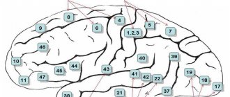
Nerve centers of the cerebral cortex
Areas of the cerebral cortex that have characteristic cytoarchitectonics and nerve connections involved in performing certain functions are nerve centers. Damage to such areas of the cortex manifests itself in the loss of their inherent functions. The nerve centers of the cloak can be divided into projection and associative.
Projection centers of the cerebral cortex are areas that represent the cortical part of the analyzer and have a direct morphofunctional connection through afferent or efferent nerve pathways with neurons of the subcortical centers.
Associative centers are areas of the human cerebral cortex that do not have a direct connection with subcortical formations, but are connected by a temporary two-way connection with projection centers. Associative centers play a primary role in the implementation of higher nervous activity. At present, the dynamic localization of some functions of the cerebral cortex has been clarified quite accurately. Areas of the cerebral cortex that are not projection or associative centers are involved in inter-analyzer integrative brain activity.
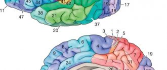
Cortical fields are functionally unequal and can be divided into primary, secondary and tertiary.
Primary fields are clearly demarcated areas that correspond to the central parts of the analyzers. The bulk of signals from the sensory organs pass into these fields along specific projection afferent pathways. Primary fields are characterized by strong development of the internal granular plate. Primary fields are associated with the relay nuclei of the thalamus and the nuclei of the geniculate bodies. They have a screen structure and, as a rule, a rigid somatotopic projection, in which individual areas of the periphery are projected into the corresponding areas of the cortex. Damage to the primary fields of the cortex is accompanied by a violation of direct perception and fine differentiation of stimuli.
The secondary fields of the cortex are adjacent to the primary fields. They can be considered as peripheral parts of cortical analyzers. These fields are associated with the association nuclei of the thalamus. When secondary fields are damaged, elementary sensations are preserved, but the ability to more complex perceptions is impaired. Secondary fields do not have clear boundaries, and the somatotopic projection is not expressed in them.
The tertiary fields of the cortex are distinguished by the finest neural structure and the predominance of associative elements. These fields are connected to the posterior nuclei of the thalamus. In the tertiary fields, the most complex interactions of analyzers are carried out, underlying the cognitive process (gnosis), and programs of purposeful actions are formed (praxia).
The cortex provides a perfect organization of animal behavior based on innate and acquired functions during ontogenesis and has the following morphofunctional features:
- Multilayer arrangement of neurons;
- Modular principle of organization;
- Somatotopic localization of receptive systems;
- Screen display, i.e. distribution of external reception on the plane of the neuronal field of the cortical end of the analyzer;
- Dependence of the level of activity on the influence of subcortical structures and reticular formation;
- Availability of representation of all functions of the underlying structures of the central nervous system;
- Cytoarchitectonic distribution into fields;
- The presence in specific projection sensory and motor systems of secondary and tertiary fields with associative functions;
- Availability of specialized associative areas;
- Dynamic localization of functions, expressed in the possibility of compensation for the functions of lost structures;
- Overlap of zones of neighboring peripheral receptive fields in the cerebral cortex;
- Possibility of long-term preservation of traces of irritation;
- Reciprocal functional relationship between excitatory and inhibitory states;
- The ability to irradiate (spread) excitation and inhibition;
- The presence of specific electrical activity.
Important VSD according to the vagotonic type