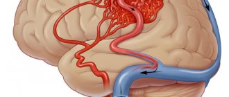
Ultrasound not only passes through tissue, but, reflected from blood cells, sends an image of the vessel to the screen, which allows one to evaluate the patency and degree of narrowing of the vessel.
There are several types of Doppler:
- Ultrasound Dopplerography (Doppler ultrasound) is a study of the vessels of the neck, head, brain or other organs, which allows you to determine the patency of the vessel, i.e. his anatomy.
- USDS - (Duplex Ultrasound Scanning) combines two functions: In this case, the vessel is already visible on the monitor, and an image of the tissue around it is obtained, as with a conventional ultrasound. It turns out that this method, unlike Doppler ultrasound, helps in diagnosing the cause of poor vessel patency. It helps to visualize plaques, blood clots, tortuosity of blood vessels, and thickening of their walls.
- With Triplex scanning, the vessel is visible on the monitor against the background of the image of the tissues in the thickness of which it passes. In this case, the vessel is painted in different colors depending on the speed of blood flow in it.
Ultrasound scanning of head and neck vessels is recommended for the following diseases:
- congenital anomalies of the location, course or branching of blood vessels
- atherosclerosis
- injury to an artery or vein
- inflammatory change in the walls of arteries and capillaries (vasculitis)
- diabetic, hypertensive, toxic angiopathy
- encephalopathy
- vegetative-vascular dystonia.
Ultrasound of the vessels of the head and neck helps to understand:
- causes of repeated transient ischemic attacks, strokes
- the degree of damage to these particular arteries due to metabolic or antiphospholipid syndromes
- the degree of impairment of the patency of blood vessels in the arterial bed due to atherosclerosis, diabetes mellitus, and smoking.
Knowledge of the condition of extra- and intracranial arteries and veins, obtained using duplex scanning, helps in prescribing the correct treatment, objective monitoring of its effectiveness, and drawing up an individual prognosis.
How the research works
Preparing for MAG duplex scanning is not difficult. It is enough not to drink strong tea, coffee, energy drinks or alcoholic drinks on the day of the examination. 2 hours before the ultrasound (and preferably longer) do not smoke. The doctor conducting the study should be warned about taking medications - they can affect the circulatory system.
Duplex scanning is an ultrasound method that is performed in two modes: standard and Doppler. Dopplerography provides the necessary data on the speed of blood flow, the direction of blood movement, and shows pathological areas. The causes of violations can be seen in the usual way using black and white images, the vessels, the blood clots and plaques present in them, and the condition of their walls are examined. If necessary, if the data after the duplex examination is insufficient, the doctor will recommend other diagnostic methods. This could be MRI or triplex vascular ultrasound, in which greater accuracy and capabilities of the method are provided by the color scanning mode.
Duplex MAG scanning is performed in the supine position. An ultrasound may include functional tests: holding or increasing breathing, finger pressure on the artery, turning the head. The procedure takes about 30-40 minutes, after which the doctor will prepare a report with the results of the examination.
Who needs to examine cerebral vessels
Duplex scanning (or at least ultrasound scanning) of intracranial arteries and veins (that is, those located in the cranial cavity) is indicated in cases of such complaints:
- headache, noise in the ears or head
- heaviness in the head
- dizziness
- visual impairment
- attacks of impaired consciousness such as fainting or inadequacy
- unsteadiness of gait
- lack of coordination
- impairment of speech production or understanding
- limb weakness
- numbness of hands.
The examination is also performed when pathology is detected during ultrasound examination of the vessels of the neck, when pathology of the neck organs is detected using CT, scintigraphy, MRI (for example, enlargement of the thyroid gland). In this case, in order to prescribe adequate therapy, a neurologist needs to know how all these diseases affect the brain, and whether its nutrition may suffer from this.
How is ultrasound examination of the vertebral and carotid arteries performed?
Before starting the procedure, the patient will be asked to remove all jewelry from the neck and free it from clothing so that the neck and supraclavicular area are freely accessible to the ultrasound probe. After this, he assumes a lying position on the couch.
The diagnostician applies a gel to the area being examined, ensuring the desired contact of the skin surface with the sensor and eliminating the presence of air bubbles that complicate the examination. It begins with determining the condition of the carotid arteries. The patient is asked to turn his head to the side and first examine the lower part of the right artery, tilting the sensor at different angles.
After this, the diagnostician leads it upward, leading it around the corner of the lower jaw. In this way, he is able to determine the depth and course of the artery, as well as identify the level at which it is divided into external and internal. The final stage is to turn on the three-dimensional color mode, thanks to which you can clearly identify areas of abnormal blood flow. The second carotid artery, as well as all its branches, are examined in the same way. Examination of the vertebral arteries involves a longitudinal placement of the sensor on the neck.
Indications for examination of the vascular bed of the head and neck
Ultrasound duplex scanning of those arteries and veins that supply blood to the brain, but are located in the neck (that is, extracranial - outside the cranial cavity) should be carried out in the following cases:
- headache
- dizziness
- unsteadiness of gait
- impaired memory, attention
- coordination problems
- when planning operations on blood vessels and heart muscles
- when identifying pathology of the neck organs, due to which the vessels passing there may be compressed
- visible contraction of the blood vessels of the heart.
Are there any contraindications?
Ultrasound waves are absolutely safe for humans, so there are no special contraindications to this type of ultrasound. The only exception is a violation of the integrity of the skin. The procedure involves contact of sensors with the head or neck, so the examination is postponed until the wound heals.
However, there are a number of special cases that make duplex examination of blood vessels difficult:
- cardiac arrhythmia, which changes the speed of blood circulation through healthy arteries and veins;
- the area under study is hidden under bone tissue;
- slow blood flow in the patient.
These are not contraindications, but in this case changes in the location of the sensors are required. It will be necessary to navigate non-standard conditions, which requires professionalism on the part of the doctor.
When is routine Doppler sonography necessary?
Doppler of both extra- and intracranial arteries and veins should be performed at least once a year as a routine study (even before the appearance of any complaints):
- all women over 45 years old
- all men over 40 years old
- those who have close relatives suffering from hypertension, diabetes, coronary artery disease
- for diabetes
- smoking
- antiphospholipid syndrome
- for osteochondrosis of the cervical spine
- metabolic syndrome
- arterial hypertension
- if you have had a stroke or transient cerebrovascular accident
- if a person suffers from rhythm disturbances (the likelihood of cerebral thromboembolism with subsequent stroke is increased)
- increased levels of cholesterol, triglycerides, low-density lipoproteins in the blood (signs of atherosclerosis)
- had surgery on the spinal cord or brain
- before planned heart surgery.
What the study will show
Duplex scanning is based on the ability of an ultrasonic wave to be reflected from moving red blood cells. This is how a visual image of the vessels is formed. The objects of study are brachycephalic vessels: carotid, subclavian, vertebral arteries and intracranial vessels.
Duplex scanning of the great vessels of the head (MAG) allows you to see and evaluate in quite detail:
- The structure of the vascular network.
- The condition of large vessels and their walls - the size of the lumen in them, the presence of atherosclerotic plaques, blood clots, the elasticity of the walls, their thickening, stratification, stretching and other deformations.
- Type of pathology, place of its occurrence and other parameters.
This information will help identify symptoms of vascular pathologies, and with timely treatment, prevent cerebrovascular accidents and such acute and dangerous manifestations as stroke.
Ultrasound scanning of the vessels of the lower extremities
A doctor may recommend ultrasound scanning of the vessels of the lower extremities for the following diseases and conditions:
- Atherosclerosis, endarteritis and diabetic angiopathy of the vessels of the lower extremities
- Atherosclerosis of the visceral branches of the abdominal aorta (vessels supplying the gastrointestinal tract, liver, spleen and kidneys)
- Aneurysm of the abdominal aorta and other vessels
- Varicose veins of the lower extremities
- Vasculitis (inflammatory vascular disease)
- Vascular diseases of the brain and neck
- Control of performed surgical intervention on blood vessels
- Postthrombophlebitic disease
- External vessel compression syndrome
- Screening examination (a study to identify asymptomatic forms of the disease)
- Thrombophlebitis and phlebothrombosis of the veins of the extremities
- Thrombosis of intestinal vessels
- Vascular trauma and its consequences
Advantages of duplex scanning of blood vessels in
Ultrasound examinations at the medical center in Maryina Roshcha are carried out by qualified doctors with extensive experience. Our doctors constantly improve their knowledge and master new diagnostic technologies. Examinations are carried out using certified modern devices from the world's leading medical equipment manufacturers. We apply an individual approach to each patient in order to quickly diagnose and provide timely assistance.
You can find out prices for duplex scanning of cerebral vessels on the website or by calling the administrators of the Diamed clinic in Maryina Roshcha.
What can she show?
Duplex scanning allows you to determine areas of narrowing of the lumen of blood vessels, the direction of blood flow, its speed, as well as the condition of the vascular walls. This study can show the location of cholesterol plaques and the presence of blood clots, which is very important when diagnosing atherosclerosis.
A decrease in the elasticity of the walls of the arteries, as well as their thickening, indicate that the patient has hypertension (high blood pressure). If a change in blood flow is diagnosed, this indicates the presence of obstacles. For example, an aneurysm is a sac-like protrusion of a vessel.
What pathologies can be diagnosed?
Doppler ultrasound has certain limitations in diagnosing diseases. For example, she will not be able to study small veins - for this she needs to perform angiography. But there are a number of pathologies that can be identified:
- Atherosclerosis. By examining the head and neck using ultrasound, it is most often possible to diagnose atherosclerotic disorders. This condition is indicated by echogenicity, the presence of plaques, and thickening of the vascular walls. Moreover, if the lumen of the artery narrows by more than 20%, then the patient is diagnosed with “stenosis.”
- Thrombosis. Thrombosis is characterized by blocking of the arterial lumen by a blood clot. On Doppler ultrasound, inflammation of the vessel wall with a homogeneous formation inside is clearly visible.
- Compression of blood vessels. The examination allows you to see areas where blood flow is impaired due to the impact of swollen tissue on the channels. This allows you to diagnose cervical osteochondrosis and identify problems with blood circulation after injuries.
In addition, these ultrasound examinations can detect a number of other diseases, including vasculitis, post-traumatic dissections of the arterial walls and other pathologies.
Doppler ultrasound is a safe diagnostic method that can be performed in any case if there is a medical need for it. Timely implementation of the procedure allows you to identify the disease at an early stage of its development.
Price policy
The cost of a duplex depends on:
- profile of the clinic;
- location;
- rating;
- personnel qualifications;
- technical equipment of the diagnostic room.
The accuracy of diagnosis and timely detection of even minor injuries depend on the quality and experience of the doctor. Do not save on duplex scanning of the head and neck, even if the cost may vary, we guarantee the result and quality of the examination performed.
If necessary, the cost of the study can include a consultation with a neurologist, phlebologist and other specialized specialists. Our medical center has the best doctors, and we also train and improve the qualifications of staff from other clinics.
In our clinic you will receive one of the best services and high quality research. You can also get treatment here. The cost of duplex scanning of the head and neck can be found in the “Price List” section. The service is provided by appointment and 100% prepayment.
Features of the method
Duplex of vessels located in the head is a study that uses ultrasonic waves, which determines the principle of operation of the equipment. Any environment (structures, tissues and elements of the body) is an obstacle to the propagation of ultrasonic vibrations. The effect is called acoustic resistance. An ultrasonic wave encounters an object, partially continues to move further with low resistance, and is partially reflected when the medium under study is significantly dense and elastic. Ultrasound is reflected from objects of the circulatory system with different intensities and speeds of reverse movement. The higher the intensity of the reflected signal, the brighter and more distinct the area of the tissue being studied appears on the screen. The reflected signal is interpreted and sent to the screen, where the doctor sees in real time the features of the structure and functioning of the blood flow system. Scanning at the extracranial level involves studying the branches of the main arteries that lie outside the cranium, but supply blood to the brain structures of the head. Examination of intracranial arteries allows us to identify disorders in the circulatory system of the brain.
Common advantages and disadvantages of the presented method
Duplex scanning has a number of obvious advantages:
- is highly informative;
- does not require the use of ionizing radiation;
- a cheaper and more accessible research method than alternative ones;
- carried out in real time, does not require waiting for a conclusion;
- helps to reflect the condition of soft tissues that are not sufficiently visualized on x-rays;
- non-invasive, performed without contrast (to which allergies are sometimes found), does not bring any unpleasant sensations;
- Suitable for children and pregnant women.
Any method, along with positive ones, also has negative sides, and in some cases it makes sense to choose other types of research. The decision is made by the doctor on an individual basis, after evaluating the results of other diagnostic procedures and tests, examination, and questioning of the patient.
The disadvantages of the method include:
- difficulty in examining small vessels;
- the possibility of diagnosis only during the research process, and not after it based on the results;
- narrow area to be examined because the bones of the adult skull impede the passage of ultrasound.
An important role in the quality of the diagnostics performed is played by the level of equipment and the professionalism of the doctor. This is one of the diagnostic methods to which increased demands are placed on the professionalism of doctors, so you should choose a clinic for duplex scanning especially carefully, because the result can differ significantly.