Find out more about diseases starting with the letter “K”: Causalgia, Brain cyst, Cluster headache, Tick-borne encephalitis, Kozhevnikov epilepsy, Colloid cyst of the third ventricle, Coma, Compressive myelopathy, Balo concentric sclerosis, Radicular syndrome, Corticobasal degeneration, Craniovertebral anomalies, Craniospinal tumor, Craniopharyngioma, Myasthenia gravis crises, Hemorrhage into the ventricles of the brain.
Depending on the location of the formation, focal symptoms may be present. The diagnosis is made based on data from computed tomography and magnetic resonance imaging of the head or neurosonography (in children). Therapy consists of aspiration and surgical removal of the formation if complications develop or its growth progresses.
General information about pathology
A cyst is a fluid-filled cavity located in the substance of the brain or its membranes. When the pathology is small in size, it has a subclinical course and is diagnosed accidentally during a neuroimaging examination of the head. Since the intracranial space has limited dimensions, with a significant increase in the volume of the formation, intracranial hypertension develops.
The size of the cavity with liquid is largely determined by the compensatory capabilities of the formation and its localization. Due to the pliability of the skull bones in children at an early age, the cyst may not manifest itself for a long time.
The formation can be found in people of different ages, both in infants and in elderly patients. Even if the fluid cavity is congenital, it can make itself felt only by the age of 30-50.
According to generally accepted practice, treatment is prescribed only in the case of a pronounced clinical picture and when complications develop. If the cavity with fluid is frozen or slowly progressing and its volumes are insignificant, a wait-and-see approach is chosen, which involves regular monitoring of the patient.
Special diet
During treatment and in the postoperative period, the patient adheres to a special diet. To reduce cholesterol in the blood, which can cause a blood clot, dishes with fatty, fried meat are removed from his diet. Meat products suitable for consumption in this situation are boiled fish, chicken and veal.
To normalize blood pressure and strengthen the immune system, fresh fruits and vegetables are included in the diet. The patient's diet is adjusted so that he eats 6-7 times a day, but in small portions. This reduces the load on the stomach, but allows all the beneficial substances from food to be fully absorbed into the intestines.
Drinking coffee and alcoholic beverages is strictly prohibited.
Classification
Classification by location:
- Cerebral (intracerebral). It is formed in areas of dead brain tissue in the internal structures of the brain.
- Arachnoid. It is formed as a result of the accumulation of cerebrospinal fluid in places of adhesions formed as a result of inflammatory processes and in places of their congenital duplication. The advantage is localized in the meninges.
The following types of brain cysts are distinguished separately:
- dermoid;
- colloidal;
- choroid plexus;
- pineal gland.
According to its genesis, a cavity with fluid can be congenital or acquired. In turn, congenital is divided into colloid and dermoid, and due to its formation into post-infectious, post-traumatic, post-stroke and echinococcal.
Identification of the disease
Modern medicine makes it possible to identify lacunar pathology. The pictures taken later clearly show the location of the tumor. They will also indicate the areas of the brain that are affected by the disease and will help determine the shape and size of the cyst. Methods used by specialists:
- head biopsy;
- blood tests for the presence of pathogenic microbes;
- blood tests for coagulation and cholesterol;
- blood pressure monitoring for 24 hours;
- MRI;
- electrocardiography;
- spiral CT;
- Dopplerography.
The principle of Doppler ultrasound
CT and MRI show the location of the tumor, the volume and affected area of the brain. Electrocardiography and Dopplerography will reflect the condition of blood vessels, arteries, the presence of heart failure and blood circulation speed. Using a biopsy, specialists will determine the type of tumor. All kinds of tests will show the condition of the blood vessels and the presence of infection. Blood pressure monitoring will also confirm pathology in the brain.
Etiology and pathogenesis
The causes of a congenital cyst are unfavorable factors that occur during the development of the fetus. These are:
- fetal hypoxia during delivery;
- fetoplacental insufficiency;
- taking a specific group of medications by a pregnant woman;
- Rh conflict between mother and child;
- intrauterine infections.
Factors that provoke the development of a congenital cavity with fluid are drug, alcohol or nicotine addiction of the mother. In this case, the child’s development takes place under conditions of intrauterine intoxication, which negatively affects the brain structures. The causes of the cavity can also be chronic decompensated diseases of the expectant mother.
An acquired cavity with fluid in the head develops as a result of:
- inflammatory diseases (encephalitis, brain abscess, arachnoiditis, meningitis);
- traumatic brain injuries;
- injuries of newborns received during childbirth;
- cerebrovascular accidents (subarachnoid hemorrhage, ischemic stroke, hemorrhagic stroke).
Depending on the etiology, the following types of liquid cavities are distinguished:
- A cyst of iatrogenic origin forms as a complication after brain surgery.
- Parasitic - develops with paragonimiasis, cerebral form of taeniasis and echinococcosis.
A cavity with fluid can also replace cerebral tissue during degenerative and dystrophic processes in the head.
If a cyst is present, there are a number of factors that can trigger its growth. These include obstruction of venous outflow from the skull, strokes and other vascular disorders, as well as head injuries, hydrocephalus, and neuroinfections.
Forms
Experts distinguish two forms of cysts: true and false. They differ in location, appearance and other characteristics.
True
It is formed in close proximity to the outer cortex of the cerebral hemisphere, but rarely affects it. The neoplasm is capable of connecting to the ventricles of both hemispheres.
Dimensions for surgery to remove Baker's cyst of the knee joint
Not all patients require removal of a knee cyst.
The size of the tumors varies from a few millimeters to 5 centimeters in diameter. In some cases, they are able to fill almost the entire space of the organ hemisphere.
The inner surface of cysts is always smooth and similar in appearance to the epindema layer. When connected to the ventricles, the formation contains cerebrospinal fluid. In other cases, the exudate is yellow in color and a large amount of protein is present.
When examined under a microscope, the surface of the cyst has altered areas, the appearance of which is caused by the incorrect structure of the cerebral cortex. The presence of hemorrhages and necrosis is established.
False
A porencephalic cyst is located in the tissues of the brain, but does not reach the cortex of the organ and does not connect to its ventricles.
Neoplasms are localized only in one hemisphere or in the white matter. A distinctive feature of false tumors is their ability to merge with each other. Single cysts are also detected.
With false neoplasms, deformation of the gyri does not occur, but scar tissue begins to form on the surface of the tumor.
Scientists have identified the bladder brain as a separate form. This is a condition in which the lesion affects both hemispheres at once, and the pathological process spreads to the brain stem.
Brain cyst
The growth of the formation at the initial stage is in most cases accompanied by symptoms of intracranial hypertension. Patients constantly complain of nausea, which has nothing to do with food, deterioration in general health and decreased performance, constant cephalalgia and pressure on the eyeballs.
In some cases, the main symptoms include a constant feeling of pulsation in the head, sleep disturbance, mild hearing loss, dizziness, motor dysfunction, fainting and tremors of the limbs. Possible visual impairment, namely double vision, visual hallucinations, deterioration of visual acuity. With high intracranial hypertension, the patient is worried about constant vomiting.
There are cases when the first signs indicating a cavity with fluid are the first occurrence of epileptic paroxysm. Subsequently, epileptic seizures recur. Paroxysms can take the form of focal Jacksonian epilepsy or absence seizures and be of a primary generalized nature.
Compared to general cerebral manifestations, focal symptoms are observed in fewer cases. These may be sensory disorders, monoparesis and hemiparesis, brainstem symptoms. The latter include dysarthria, eye movement disorders, and swallowing disorders.
One of the complications of formation is cyst rupture. In this case, hemorrhage due to rupture of the vessel, compression of the brain, the formation of an epileptogenic focus and occlusive hydrocephalus are possible.
In the congenital form of the cyst, episyndromes and intracranial hypertension are recorded at an early age. Education in the brain can cause the child to develop mental retardation and mental development disorders.
Clinical picture
Symptoms of the development of such a formation may not appear for years. In such patients, cysts are detected during routine head examinations.
As the tumor grows, it compresses nearby parts of the brain. Symptoms that indicate a lacunar cyst depend on the location of the affected areas:
- For example, if a cyst has formed in the basal ganglia area
, the patient will complain of problems performing complex movements. - When the parietal lobe
is damaged, coordination of movements, a sense of balance and body position in space are impaired, and oral and written speech skills regress. - If the formation is located in the temporal region
, auditory, olfactory, and taste hallucinations may occur. - The formation in the frontal part
causes involuntary facial movements and epileptic attacks. - Impaired functioning of the occipital region
leads to vision problems.
Also characteristic symptoms indicating damage to the membranes of the brain are nausea, vomiting, headaches, photophobia, and loss of elasticity of the neck muscles.
Types of brain cysts and their symptoms
Arachnoid - occurs in almost 4% of the population. A cavity with fluid can be congenital or acquired. In the latter case, it develops in response to traumatic brain injury. The formation is localized on the surface of the brain in its membranes. The cavity is filled with cerebrospinal fluid.
In most cases, an arachnoid cyst does not make itself felt for quite a long time and is discovered by chance. Severe symptoms appear only if a large amount of fluid accumulates in the cavity. In this case, cerebrospinal fluid is produced by the cells lining the cavity.
With a sharp increase in the volume of the cavity with liquid, it can rupture and, as a result, die.
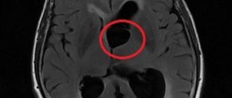
Colloid cysts are recorded in 15-20% of all cases of formations inside the ventricles of the brain. Most often it is localized above the foramen of Monroe in the anterior region of the 3rd ventricle. Less common in the area of the transparent cerebral septum in the 4th ventricle.
The liquid filling the cavity of the colloid cyst has high viscosity. Patients experience symptoms of hydrocephalus and, with a certain position of the head, a paroxysmal increase in cephalgia is noted.
In rare cases, memory loss, behavioral disorders and weakness in the limbs occur.
Pineal cyst of the pineal gland - according to statistics, 10% of patients have formations of this type that are small in size and do not make themselves felt. Cystic formations are localized in the epiphysis of the brain, in most cases they are no more than 1 cm in size. Otherwise, symptoms appear. When it grows, it can block the entrance to the “plumbing” of the brain and block the circulation of cerebrospinal fluid, causing occlusive hydrocephalus.
Epidermoid or dermoid is a formation that is an anomaly of intrauterine development. In this case, the cells of the baby's future skin and its appendages remain inside the brain. Accordingly, in addition to liquid, elements of the ectoderm are present, namely sebaceous glands and hair follicles.
After the birth of a child, such a cyst quickly increases in size. The only possible treatment is surgical removal of the formation.
Choroid plexus cyst - forms regardless of the person’s age. In this case, the space between the plexus vessels is filled with cerebrospinal fluid. Symptoms are rarely present, sometimes accompanied by epileptic seizures and symptoms of intracranial hypertension.
With a congenital choroid plexus cyst, the formation is diagnosed at the 20th week of intrauterine development using ultrasound. By the 28th week, such formations resolve.
Traditional medicine treatment
Despite the seriousness of the disease, lacunar cysts have several methods of treatment with folk remedies. In general, it, like conservative therapy, is aimed at eliminating the causes of the tumor. Therefore, traditional methods must be used in combination with drug therapy and only with the permission of the attending physician.
Medicinal plants that can positively affect a person’s condition in this situation are hemlock, elecampane, wormwood, chamomile, calendula, yarrow, raspberry, corn silk, and Dioscorea Caucasica.
From these plants you can make decoctions or alcohol tinctures. The decoction is simple to make - 1 tbsp. a spoonful of the plant is poured into a glass of boiling water and simmered over low heat for no more than 15 minutes. After the broth has cooled, it should be strained and taken in a glass half an hour before meals.
The alcohol infusion takes longer to make - the dry, crushed plant is filled with alcohol in a ratio of 1 to 3, that is, 300 ml of alcohol per 100 g of plant, and infused for 2 months in a dark place. The infusion should be shaken once a week. After straining, the product is taken 1 teaspoon per day, half an hour before meals.
Diagnostic measures
A neurologist may suspect the presence of an intracranial formation of significant size based on the patient’s neurological status and clinical symptoms. In this case, the patient is sent for examination to an ophthalmologist and otolaryngologist to check vision and hearing. Specialists perform ophthalmoscopy, audiometry, perimetry and visometry. With severe hydrocephalus, ophthalmoscopy reveals congested optic discs.
By referring the patient for echo-encephalography, it is possible to diagnose increased intracranial pressure. If the patient experiences epileptic paroxysms, he is additionally sent for electroencephalography.
It is extremely important to differentiate a cavity with fluid from a tumor, abscess and hematoma. It is not possible to do this on the basis of collected clinical data alone. Therefore, to make a clear diagnosis, neurologists use neuroimaging diagnostic methods.
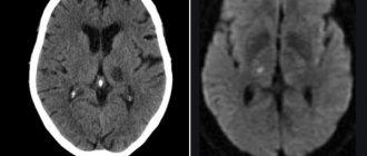
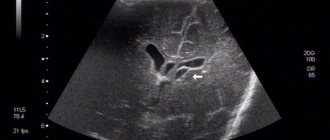
By performing an ultrasound examination, it is possible to diagnose certain types of congenital cysts even at the stage of intrauterine development of the fetus. After birth, until the baby's large fontanelle closes, neurosonography is performed to make the correct diagnosis. In adulthood, to visualize a brain cyst, the patient is sent to a magnetic resonance or computed tomography scan of the head.
MRI and CT are performed with contrast in order to differentiate a cyst from a tumor. A cavity with liquid is not able to accumulate a contrast agent, unlike a tumor.
After diagnosis, constant monitoring of the patient with a cystic formation is important. During regular examinations, the doctor monitors the volume of the cyst over time.
If the cyst is a consequence of a stroke, additional vascular examinations are carried out: ultrasound, MRI and CT of vessels, duplex scanning.
General principles of therapy
Drug therapy for cystic formations has practically no results. The only possible treatment is surgical removal of the fluid cavity. But, most forms are small in size and remain in a “dormant” state for many years. In this case, no methods of therapy are used; a wait-and-see regimen is chosen and the patient is regularly examined.
Formations that are accompanied by symptoms of hydrocephalus, complicated by bleeding and rupture, compressing the brain and rapidly increasing in size must be removed. Only a neurosurgeon decides which method of surgical treatment to use.
If the patient has a disorder of consciousness (coma or stupor), he is urgently referred to external ventricular drainage. This method allows you to reduce compression of the brain by the cyst and intracranial pressure. If the cyst ruptures or hemorrhages, surgery is performed. The patient undergoes craniotomy and the formation is excised.
In the absence of complications and disturbances of consciousness, the operation is performed planned and endoscopically. The advantage is the patient’s quick recovery period and low trauma. With endoscopic access, a milling hole is made in the skull, through which fluid is sucked out from the cavity. In order to avoid subsequent accumulation of fluid, several holes are made in the cavity (connected to the cerebrospinal fluid space) or cystoperiotoneal shunting is performed (a special shunt is installed).
The postoperative period involves rehabilitation therapy, including exercise therapy, reflexology and massage. The patient is prescribed drugs to improve blood supply to the brain, absorbents and decongestants.
Surgery
If the cyst poses a danger to the patient’s condition, then surgery is indicated. It can be performed using various methods - bypass surgery, endoscopy and craniotomy.
Each method has both positive and negative sides. Endoscopy can only reach those cysts that are located directly under the skull bone. The instrument, which is a thin tube, does not reach the neoplasms lying deep in the brain matter.
Bypassing a cyst involves suctioning out the fluid filling the cyst using a thin needle. As a result of this procedure, the cyst is reduced until only one shell remains. The negative side of this procedure is the risk of introducing an infection into the brain, which will cause complications.
Craniotomy involves opening the skull to provide access to the cyst at any depth. This type of operation provides a 100% opportunity to completely remove the tumor, but has a long postoperative period.
The type of operation is chosen by the attending physician, based on the patient’s condition, location and size of the cyst, and many other medical indicators. The wishes of the patient in this situation are not taken into account.
Forecasts
In most cases, a frozen cavity with liquid of insignificant size does not bother the patient and does not cause any symptoms. In other cases, with adequate and timely treatment, the outcome is favorable.
In rare cases, patients after surgical removal of a cyst experience a residual moderate-to-severe liquor-hypertensive symptom. If a focal neurological deficit develops, it persists after treatment.
With the removal of the cyst, epileptic paroxysms disappear, but often appear later. This is justified by changes in the operated area of the head, in particular the formed adhesions. Secondary epilepsy is practically uncontrollable by anticonvulsant therapy.
Consequences
The lack of quality therapy leads to the worsening of old symptoms and the appearance of new ones. A person loses coordination, his vision decreases, up to complete loss. Facial expressions and speech deteriorate, and it becomes difficult to walk. A person can be paralyzed.
Infants are at risk of developing hydrocephalus. Liquor fluid accumulates in the ventricles, and the brain contracts. With proper diagnosis and treatment, you can get rid of symptoms and not worry about the appearance of new formations. The main thing is a correct approach to the problem and prevention, which usually lasts a little less than 2 years.
A lacunar cyst is a benign tumor that does not develop into a malignant one. If the disease is not treated, the formation will increase in volume and affect parts of the cerebral cortex, impairing the functioning of the body. There is no need to panic; in most cases, the cyst does not interfere with the functioning of organs and systems, and the person lives peacefully, unaware of the pathology.
Prevention
An acquired cyst most often becomes a consequence of developing inflammatory, vascular, infectious and post-traumatic processes. Therefore, it is absolutely obvious that only correct and timely treatment of any pathologies, including resorption and neuroprotective therapy, can serve as a preventive measure for the development of formations in the brain.
The only measure to prevent congenital cysts is to protect the woman and fetus from exposure to provoking factors. Equally important are correct management of pregnancy and delivery.