Dislocation syndrome reflects a pathological process that develops as a result of displacement of brain structures relative to each other. This is a complex of clinical, pathomorphological, pathophysiological signs that manifest a shift in the hemispheres or cerebellum. The progression of the pathology leads to damage to the trunk, which is accompanied by hemorrhagic, ischemic, atrophic changes, which often causes the death of the patient.
Definition of pathology
Dislocation syndrome is the process of moving a fragment of brain tissue relative to other structural elements of the skull and brain. The skull is the hard part of the head. As the volume of the contents of the cranium increases, nerve tissue may bulge through the natural openings at its base. Supporting partitions that divide the contents of the skull into sections limit the possibilities of compensation for pathological displacement of brain fragments.
The brain inside the cranium does not occupy the entire volume. Between the medulla and the arachnoid membrane is the subarachnoid space. In areas of expansion of the subarachnoid space, subarachnoid cisterns are formed. As a result of various pathological processes, areas of distension (pressure) may appear in certain parts of the brain, which provokes a displacement of brain structures.
Pathological processes differ in etiology, but they cause the same shift in brain regions. Dislocation syndrome of brain structures manifests itself with the same type of clinical picture. Symptoms when there is a shift in parts of the brain do not depend on the nature of the pathological process, but depend on the localization of the lesion, the rate of development of the disorders and the volume of the structures involved. Dislocation syndromes are protrusions of the brain matter into the natural openings of the skull and open spaces of the dura mater.
How does this happen
The GM does not occupy the entire volume of the skull. Between it and the bone walls there is a space in which there are three meninges that provide physical protection and nutrition. When a factor appears that increases pressure inside the skull, the sections shift to free places where the subarachnoid space is located.
As a result of dislocation, hernias are formed - areas in the head that protrude into the cracks and openings of the skull. The presence of a cerebral hernia in itself does not lead to serious disorders. The danger lies in a complication - pinched cerebral protrusion - a condition in which the displaced area of the brain stops receiving blood and dies.
There are two types of offsets:
- Lateral dislocation of the brain
- Axial dislocation (axial displacement).
Classification of forms and types
Dislocation of areas of the brain is a pathology that occurs with different intensities, which suggests division into types depending on the degree of displacement of the substance:
- Bulging. Slight displacement relative to natural location.
- Wedging. A protrusion of brain tissue past fixed structures, for example through openings in the skull.
- Infringement. Accompanied by a cessation of blood supply to the area due to compression of the blood vessels.
Depending on the direction of displacement, axial (relative to the axis of the trunk) and lateral forms are distinguished. In medical practice, there are types of dislocation of brain structures:
- Displacement of hemispheric structures under the area of the falciform process in the dura mater.
- Displacement of the temporotentorial direction.
- Shift of the cerebellar-tentorial direction.
- Shift of the cerebellar tonsils into the foramen magnum.
- Moving the bridge through the hole located in the tentorium of the cerebellum.
- Moving into the space of the side tanks in the bridge area.
- Shift of the corpus callosum into the cistern in the dorsal direction.
- Movement of the substance of the frontal lobe into the cistern in the area of the chiasm.
- External type dislocation (bulging of the brain matter through a hole formed in the skull as a result of injury).
The first 4 variants of dislocation syndrome are of particular importance in neurology, because they can provoke a significant deterioration in the patient’s condition and death. Axial dislocation of the brain substance is a transtentorial herniation - the temporal lobe (medial part) protrudes through the cerebellar tentorium (supports the temporal lobe) and partially under it.
Axial dislocation leads to compression of the ipsilateral and contralateral nerves, the posterior artery, and as it progresses, damage occurs to the brain stem and areas surrounding the thalamus. Shift of the medulla under the falciform process occurs when the angular gyrus moves under the falx cerebri (as a result of pressure on it from an increasing volume lesion in the hemisphere).
As a result, the anterior arteries are compressed, which provokes a heart attack localized in the paramedian cortex. As the area of infarction increases, the risk of developing central or transtentorial herniation increases. Central dislocation of brain structures occurs as a result of tumor processes of bilateral localization or diffuse brain edema. In this case, the temporal lobes bulge through the notch (recess) of the cerebellar tentorium.
Protrusion of the cerebellar tonsils (“small brain”) is most often associated with a volumetric intracranial formation of infratentorial localization. The shift of the cerebellar tonsils through the foramen magnum is accompanied by compression of parts of the trunk, which leads to blocking the outflow of cerebrospinal fluid.
Causes
Regardless of the etiology of the primary pathological process, a shift in the medulla is always associated with compression and mechanical damage to tissue, resulting in edema and hydrocephalic syndrome. The main reasons for the development of edema and dislocation of brain structures:
- Space-occupying formations in the cranial cavity - tumors, cysts, areas of hemorrhage (compression-dislocation syndrome develops).
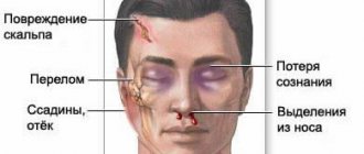
- Traumatic brain injuries.
- Hydrocephalus, increased intracranial pressure (hypertension-dislocation syndrome develops).
- Infectious diseases of the central nervous system.
Compression and subsequent displacement of the medulla occurs due to edema and mechanical impact from the volumetric intracranial formation. Edema develops due to impaired circulation of blood and cerebrospinal fluid. The displaced areas of the brain additionally cause compression of the blood vessels and cerebrospinal fluid circulation pathways, which aggravates the progression of edema.
Data decryption
h24,0,0,0,0–> p, blockquote17,0,0,0,0–>
EchoEG is based on three complexes (or bursts):
p, blockquote18,0,0,0,0–>
- The initial complex is formed by sound waves reflected from the dense bone tissue of the skull, skin and superficial brain structures from the side located next to the sensor.
- The average complex is obtained when ultrasound waves collide with brain structures located between the hemispheres.
- The final complex is obtained from the soft tissue and dura mater on the opposite side of the sensor.
ECHO of the head connects these signals and, based on the received impulses, reflects them on the monitor in the form of a graphic diagram. Decoding begins with data analysis:
p, blockquote19,0,0,0,0–>
- M-echo is a signal with the highest amplitude, occupying an average position between the two complexes. Displacement up to 5 mm is allowed. In neurological disorders, a displacement of more than 6 mm is an alarming factor requiring additional diagnostics.
- A dilated or disrupted signal received from the third ventricle indicates high intracranial pressure.
- The ripple of M-echo signals should not exceed 30-40%. If the indicators are at the level of 50-70%, then this is a clear sign of hydrocephalic syndrome.
- The SI or average sellar index is within the range of 3.9-4.1. If it is low, then this indicates high intracranial pressure. This indicator is not assessed in children.
”alt=””>
p, blockquote20,0,0,1,0–>
- Hemorrhagic cerebral stroke: what it is, symptoms and consequences
Symptoms of the disease
Regardless of the etiology of the pathological process that provoked the dislocation of brain fragments, an increase in intracranial pressure values more often occurs, which is accompanied by the appearance of the Cushing reflex. The pathological reflex combines the following symptoms: an increase in arterial (systolic, top number) and pulse (the difference between systolic and diastolic pressure) pressure, respiratory dysfunction, bradycardia (disturbance of the sinus rhythm of the heart).
Brain dislocation syndrome manifests itself depending on the type of displaced structures and direction. If the medulla moves under the falx process, the following symptoms are observed:
- Hemiparesis (paresis in one half of the body).
- Hemihypesthesia (loss of sensation in one half of the body).
- Pathological reflexes (grasping, oral automatism).
- Hemiataxia (impaired motor coordination in one half of the body).
- Mental disorders characteristic of damage to the frontal lobe (blurred consciousness, memory impairment, disorientation).
- Hyperkinesis (pathological involuntary movements).
In the axial form, signs appear at an early stage: hyperthermia (overheating of the body), tachycardia (rapid heartbeat), flickering consciousness (alternating periods of conscious and unconscious states), hypercatabolism (increased catabolism involving tissue proteins), apnea (short-term cessation of breathing), narrowing pupils. There is also an increase in muscle tone.
The late stage is manifested by loss of consciousness, coma, immobility, lack of response to painful stimuli, and hormetonic type convulsions (cyclical, rhythmic spasms). Progression of the pathological process can lead to the development of coma and terminal condition, death. Clinic of dislocation syndrome with temporal lobe shift:
- Pupil dilation (unilateral).
- Weakening of the pupil's reaction to a light stimulus.
- Gaze paresis.
- Hemiparesis (paresis in one half of the body).
- Hemiplegia (loss of the ability to make voluntary movements).
- Hyperthermia (body overheating, excessive heat accumulation).
- Bradycardia (disturbance of sinus rhythm of the heart).
The progression of the pathological process is accompanied by the development of stupor, coma, and severe oculomotor disorders. If left untreated, a terminal condition develops. When there is a herniation into the foramen magnum, symptoms (rigidity and pain in the occipital muscles, cerebellar disorders, bradycardia, bulbar disorders) usually increase rapidly. Often there is a rapid fatal cessation of cerebral circulation and respiratory arrest. Due to the high rate of progression of this type of pathology, morphological changes are not of great importance.
Clinical manifestations of brain dislocation in the acute period of severe traumatic brain injury
The results of surgical treatment of 168 patients with dislocation syndrome in acute severe traumatic brain injury were treated in the National Hospital. The analysis of the clinical manifestations and treatment of dislocation of the brain in patients with severe traumatic brain injury in the acute period. The dynamic observation of dislocation syndrome clinic patients examined. Outcomes depend both on the primary and secondary factors of brain damage.
Key words: Severe craniocerebral trauma, dislocation syndrome, a horizontal and axial dislocation, surgical treatment.
Relevance of the problem : In victims with severe traumatic brain injury (STBI), intracranial hypertension and dislocation manifestation of the brain structure are an important link in the pathogenesis of this nosology, and is the main cause of mortality [3, 9, 20].
According to research by L. B. Likhterman (1999), mortality in severe TBI in the Russian Federation is at least 68–70%, and, according to S. V. Astrakov (2007), from 38 to 80%.
Dislocation syndrome (DS) is a common complication of emergency neurosurgical diseases. As reserve cerebrospinal fluid spaces are exhausted, various intracranial pressure gradients may arise. This leads to displacement of brain structures, deformation and infringement of the brain [3,6]. Dislocation syndrome with STBI develops in a short period of time, resulting in an immediate threat to the patient’s life requiring emergency medical care [4, 8].
The clinical picture of acute dislocation syndrome does not depend on the etiology of the process. The clinical manifestation of brain dislocation in different patients depends on the rate of development of the dislocation, localization and volume of the pathological process of the brain [1, 6, 18].
The critical level of increase in intracranial pressure is considered to be 20–25 mmHg. Art., although clinical experience demonstrates the absence of a close connection between the absolute value of intracranial pressure and the development of brain dislocation. However, in the presence of a volumetric process of temporal localization, dislocation and herniation can occur even with intracranial pressure less than 20 mm Hg. Art. [13]. Intracranial pressure level is more than 20–25 mmHg. Art. in most cases, it requires the appointment of adequate anti-edematous therapy, and in some cases, surgical decompression.
Treatment of brain injuries accompanied by dislocation syndromes is an extremely complex, at the same time unsolved to date [2] and controversial problem.
According to most retrospective and prospective non-randomized clinical studies, hemicraniectomy with dural plastic surgery reduces associated mortality from 80 to 30% [15, 16, 19]. Some studies indicate that mortality may be further reduced if hemicraniectomy is performed within the first 24 hours after stroke [14].
All neurosurgeons write about the severe course of dislocation syndrome and the danger to the patient’s life. However, recently little attention has been paid to the treatment, including surgery, of this formidable complication. And only a few reports describe attempts to eliminate dislocations [5,10,11,12,18].
Purpose of the study: To analyze the clinical manifestations of brain dislocation in the acute period of severe traumatic brain injury, depending on the location, form and degree of manifestation of brain dislocation and to develop principles for eliminating brain dislocation.
Research objectives:
- To determine the location and types of the source of damage in the acute period of TBI and, depending on the severity of the brain contusion, to establish the types and forms of dislocation syndrome.
- To study the clinical manifestation of dislocation syndrome with various forms and degrees of brain dislocation based on neuroimaging examination methods.
- To analyze comparative results in surgical and conservative treatment of brain dislocations.
Materials and methods: The results of clinical and instrumental examination methods, as well as surgical treatment of 168 patients with severe traumatic brain injury (STBI), who were hospitalized in the neurosurgery clinic of the National Hospital of the Ministry of Health of the Kyrgyz Republic, aged from 21 to 61 years, were studied. , on average 40–41 years old. 138 (82.1%) patients were men, 30 (17.9%) patients were women.
To assess the degree of dislocation syndrome (DS), the following signs were taken into account: the level of depression of consciousness [7], general cerebral and focal symptoms, respiratory rate, pulse, blood pressure. Instrumental examination included CT and (or) MRI of the brain. Consciousness of patients was assessed using the Glasgow Coma Outcome Scale [17]. In most cases (76%), the operation was performed by decompressive craniotomy with removal of the lesion and elimination of dislocation; in other cases, resection craniotomy or osteoplastic craniotomy.
In most cases, patients 137 (81.5%) were transported by ambulance after injury from 30 minutes to 12–14 hours, on average 6–7 hours. In the remaining cases (18.5%), patients were admitted to the nearest medical institutions, and after first aid was provided, they were subsequently transferred to a neurosurgical clinic.
There were 112 patients with closed craniocerebral injury, 56 patients with open craniocerebral injury, of which 7 cases had penetrating TBI. Various types of fractures of the vault and base of the skull were found in 97 patients.
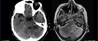
CT examination was performed in 84 patients, and MRI in 65 patients. 135 patients underwent surgical treatment, 33 patients received conservative treatment.
Results and discussion : Subdural hematoma (SDH) was found in 42 patients, epidural hematoma (EDH) in 19 patients, a focus of contusion with hemorrhagic impregnation and the formation of intracerebral hematoma in 24 patients, sub- and epidural hematoma in 12 patients, in 22 cases focus of contusion with hemorrhage and epi-, subdural hematomas, multiple foci of contusion with crushing of the brain substance with hemorrhage in 8 patients. In 57 (33.9%) patients there was bilateral brain damage and in 111 (66.1%) cases a unilateral focal lesion was established (on the left in 48 patients and on the right in 63 patients).
CT and MRI examinations of the brain revealed horizontal and axial types of displacement (dislocation) of brain structures. Horizontal displacement (under the falx of the brain) was found in 49 patients, axial displacement (at the level of the tentorial foramen or foramen magnum) in 16 patients, horizontal and axial dislocations were found in 94 patients. Thus, the following forms of dislocation have been established: 1) cingular (under falx) dislocation in 49 cases; 2) temporotentorial dislocation, 14 cases; 3) temporotentorial and foramenal dislocations were found in 2 patients. 4) cingulate and temporal-tentorial dislocation were found in 78 patients. 5) cingulate, temporotentorial and foraminal dislocations in 16 patients.
Horizontal - transverse types of displacement were identified in 111 (86.6%) of 127 patients with unilateral lesions, and in 35 (85.3%) of 41 patients with bilateral lesions.
In unilateral traumatic lesions out of 127 patients, axial dislocation was observed in 75(59%) cases, in 34(46.6%) patients temporotentorial dislocation up to 4mm, in 31(41.3%) patients more than 4mm. With bilateral focal lesions, out of 41 patients, temporotentorial dislocation was observed in 29 patients, temporotentorial and foramenal dislocations were found in 2 patients. Bilateral temporotentorial dislocation was found in 3 patients.
According to our data, the degree of horizontal type of brain dislocation depended on the location of the lesion. When the lesion was localized in the parietotemporal region, out of 19 patients, in 5 (26.3%) cases, the displacement of the median structures was 10 mm or more, in the fronto-parietotemporal region, out of 31 patients, 14 (45.1%) cases, as well as with extensive traumatic damage to one hemisphere of the brain out of 17 patients in 6 (35.3%) cases. When the lesions were localized in the frontal lobe of the brain, out of 14 patients, horizontal dislocation in 6 (42.6%) patients was 4 mm, in 5 (35.7 %) patients up to 8 mm. In case of traumatic lesion of the parietal region, out of 7 patients, in 3 patients the horizontal dislocation reached 4 mm and in 1 patient up to 8 mm. With bilateral lesions out of 41 patients, a horizontal transverse dislocation with a unilateral accent of displacement was detected, but the size of the displacement was not expressed, in 12 (29.2%) patients up to 4 mm and in 15 (36.5%) up to 8 mm.
Table 1
Causes of development of dislocation syndrome ( n -168)
| Types of dislocation | Cingular dislocation | Axial dislocation | ||||||
| With one side | On both sides | |||||||
| Dimensions (mm) mixing Types of outbreak defeats | Up to 4mm | Up to 8mm | Up to 12mm | Up to 17–18mm | Up to 4mm | More than 4mm | Up to 4mm | More than 4mm |
| 5 | 4 | 6 | — | 5 | 4 | — | — |
| 8 | 9 | 15 | 1 | 2 | 9 | 1 | — |
| 6 | 8 | 5 | 1 | 7 | 5 | 1 | 1 |
| 4 | 11 | 12 | 7 | 13 | 11 | — | — |
| 2 | 1 | 5 | — | 2 | 4 | 1 | 0 |
| 12 | 15 | 8 | — | 7 | 7 | 8 | 11 |
EDH-epidural hematoma; SDH-subdural hematoma; OUiRGM VMH - foci of contusion and crushing of the bare brain, intracerebral hematoma; MOUiRGMG - multiple foci of contusion and crushing of the brain with hematomas; Combined - VMG + SDH or + EDH in one hemisphere; DMOUiRGVG - bilateral multiple foci of contusion and crushing of the brain.
Satisfactory - moderate residual effects and recovery;
Unsatisfactory - gross residual effects or vegetative state;
Axial types of dislocation were often observed in bilateral lesions out of 41 patients in 33 (80.4%) cases: bilateral temporotentorial herniation was found in 19 (46.3%) patients, 8 cases up to 4 mm and in 11 patients more than 4 mm, unilateral temporotentorial herniation. tentorial herniation in 14 (34.1%) patients, of which up to 4 mm in 7 and more than 4 mm in 7 patients. With a unilateral lesion, frequent temporotentorial dislocation was observed when the lesion was localized in the parietotemporal region out of 19 patients in 15 (78.9%) cases, temporal localization of the lesion out of 6 patients in 4 (66.6%) patients, fronto-parieto- temporal localization out of 31 patients in 21 (67.7%) patients. With extensive localization of the lesion, out of 17 patients, 14 (82.3%) cases, in the frontotemporal region, out of 12 patients, 8 (66.6%) cases.
Cingular and temporotentorial forms of dislocation were often observed with combined lesions, in 6 (70%) cases out of 8 patients, and in 24 (69%) cases with multiple lesions with hematomas out of 34 patients.
The clinical picture of 49 (29.1%) patients with the cingulate form of dislocation was marked by general cerebral symptoms, focal symptoms were detected in 31 (63.2%) patients, mild brainstem symptoms in the form of pupil dilation, absence of photoreaction and decreased corneal reflexes in 9( 18.4%) patients.
Of the 78 (46.4%) patients with cingulate and temporotentorial dislocation: in 66 (84.6%) patients, along with general cerebral symptoms, focal disorders in the form of pyramidal insufficiency dominated, in 41 (52.6%) patients the lungs stem symptoms, 14 (17.9%) patients had severe brainstem symptoms with impaired breathing, cardiac activity, decreased and lack of reactions to painful stimuli.
With temporotentorial dislocation, out of 14 (8.3%) patients: focal symptoms were observed in 9 (64.2%) patients, 3 patients had mild brainstem symptoms and 4 patients had severe brainstem symptoms.
Of 16 (20.5%) patients with cingulate, temporotentorial and foramenal dislocation: focal symptoms were detected in 7 (43.7%) patients, mild brainstem symptoms in 4 patients and severe brainstem symptoms in 6 (37.5%) patients symptoms.
In 2 patients with temporotentorial and foramenal dislocation, the condition was extremely severe with severe brainstem symptoms.
When analyzing the results of conservative treatment, we identified patients admitted at the stage of compensation according to GCS 12–14 points, displacement of the midline structures of the brain not exceeding 5 mm and the volume of the hematoma not exceeding 50 cm3. showed good results. Of the 33 patients, 13 (39.1%) were discharged with recovery (on the Karnofsky scale 70–90%), 9 (27.3%) patients were in unsatisfactory condition (on the Karnofsky scale 50–60 points). In extremely serious condition, the admitted patients had a GCS score of 3–5 points, with multiple foci of brain damage, with horizontal dislocation of more than 10 mm and axial dislocation of 2–4 mm, with severe vital disturbances, 10 (43.5%) patients, despite the intensive 6 patients died within 48 hours of therapy and 4 patients died before the 4th day.
Surgical treatment was performed in 135 patients using three methods: decompressive craniotomy (DCT) - 51 (37.8%) patients, osteoplastic craniotomy (OPCT) - 49 (36.3%) and resection craniotomy (RTC) - 35 ( 25.9%) to patients with removal of hematomas and foci of contusion and crush injuries with elimination of dislocation syndrome. Of the 135 patients operated on, 69 (51.1%) patients were discharged in satisfactory condition (on the Karnovsky scale 80–90 points) with recovery, 25 (18.5%) patients were in unsatisfactory condition (on the Karnovsky scale 50–60 points), not Despite intensive therapy, 39 (28.9%) patients died.
The outcomes depending on the location of the lesion were as follows. Multiple lesions with hemorrhage in the brain out of 8 patients, death was in 5 (62.5%) patients, 2 (25%) patients were discharged in unsatisfactory condition according to the Karnofsky scale 60–60 points, 1 (12.5%) the patient was discharged in satisfactory condition. With bilateral lesions, out of 41 patients, death occurred in 17 (41.4%) patients, unsatisfactory condition on the Karnovsky scale of 60–70 points in 12 (29.3%) patients and 12 (29.3%) patients with satisfactory results . In other cases, treatment outcomes are shown in Table 2.
table 2
Outcomes of surgical and conservative treatment
| Satisfactory | Unsatisfactory | Death | Total | |
| EDH | 15 | — | 3 | 19 |
| SDH | 29 | 4 | 9 | 42 |
| OUiRGM VMG | 16 | 3 | 5 | 24 |
| Combined | 14 | 11 | 9 | 34 |
| MOUiRGMG | 1 | 2 | 5 | 8 |
| DMOUiRG | 12 | 12 | 17 | 41 |
The outcomes depending on the degree of dislocation were as follows. In case of temporotentorial dislocation, out of 14 patients, with a fatal outcome, 6 (42.8%) patients had a displacement of the hippocampal gyrus between the free edge of the tentorium of the cerebellum and the brain stem of more than 4 mm. 5 (35.7%) patients were discharged with gross residual effects, where the hippocampal gyrus was herniated to 2–4 mm.
With a mixed type of dislocation (cingular, temporotentorial and foramenal dislocation), 15 (93.7%) of 16 patients died within 7 days after injury, 1 patient was discharged with gross residual effects. In these patients, a dislocation of the hippocampal gyrus into Bichat's fissure of more than 4 mm and a displacement of the cerebellar amygdala into the foramen magnum of up to 3–6 mm were revealed.
There were 2 patients with temporotentorial and foramenal dislocation, both with fatal outcomes, where dislocations of brain structures were also revealed in the Bichat fissure of more than 4 mm and in the foramen magnum up to 3–6 mm.
With cingular and temporotentorial dislocation, out of 78 patients, death was observed in 22 (28.2%) cases, displacement under the falx of the cingulate gyrus was up to 17 mm, and into the Bichat fissure of the hippocampal gyrus more than 4 mm. There were 22 (28.2%) patients with gross residual effects, where the dislocation under the falx reached 14 mm and in the Bichat gap up to 4 mm. With moderate residual effects and recovery in 33 (42.3%) patients, herniation under the falx was up to 12 mm and between the free edge of the tentorium of the cerebellum and the brain stem up to 2–4 mm.
Relatively good results were obtained with cingulate dislocation; out of 49 patients with a satisfactory result, 44 (89.7%) patients were discharged, where the dislocation of the cingulate gyrus under the falx was 4–8 mm.
Conclusions:
- Axial types of dislocation often occurred when the lesion was localized in the parietotemporal and fronto-parietotemporal regions. And with combined and multiple lesions, a more severe degree of dislocation developed.
- For the cingulate form of dislocation after surgical treatment, good results of up to 73.4% were observed. With temporotentorial dislocation (35.7–42.8%), treatment outcomes are unfavorable, and with temporotentorial and foramenal dislocation, a 100% fatal outcome is observed.
- Surgery is a more effective way to eliminate brain dislocation.
Literature:
- Akhmedov E. A., Kasumov R. D., Bersnev V. P. Differential tactics of surgical treatment of traumatic chronic subdural hematomas. // Polenov readings. Saint Petersburg. 2009.-36s.
- Bersnev V.P. On the issue of surgical treatment of hypertensive intracerebral hemorrhages / V.P. Bersnev, M.K. Agzamov // VI Polenov Readings. St. Petersburg, 2007. - 145 p.
- Blinkov, S. M. Displacements and deformations of the brain. Morphology and clinic /S. M. Blinkov, N. A. Smirnov. - L.: Medicine, 1967.-202 p.
- Zotov Yu. V., Kondakov E. N., Shchedrenok V. V., Kondratyev A. N. Intracranial decompression of the brain in surgery for severe traumatic brain injury. - St. Petersburg, 1999. -142 p.
- Lebedev V.V., Bykovnikov L.D. Guide to emergency neurosurgery. - M., “Medicine”, 1987.-335 p.
- Lebedev V.V., Krylov V.V. Emergency neurosurgery: A guide for doctors. M.: Medicine, 2000.-257 p.
- Konovalov A. N. Clinical guidelines for traumatic brain injury. Volume I / A. N. Konovalov, L. B. Likhterman. 1998.-549p.
- Kondakov E. N., Klimash A. V., Bakhtiyarov A. K., Bokin V. D. Supratentorial traumatic dislocation of the brain / Zh. Neurological Bulletin - 2008 vol. - XL, issue 3 - pp. 19–24.
- Potapov A. A. Modern recommendations for the diagnosis and treatment of severe traumatic brain injury // Potapov A. A., Krylov V. V., Likhterman L. B., Tsarenko S. V., Gavrilov A. G., Petrikov S. S. Zh. “Issues of neurosurgery named after. N. N. Burdenko” 2006. No. 1. P. 3–8
- Saribekyan A. S. Tactics of surgical treatment of severe head injury and traumatic intracranial hemorrhage in terms of the dynamics of intracranial hypertension: Abstract of thesis. diss. doc. medical sciences - Moscow, 1992.C - 23–24.
- Saribekyan A. S. Tentoriotomy and ventricular drainage in the surgical treatment of severe TBI: Abstract of thesis. diss. Ph.D. medical sciences - Moscow, 1984. - 26 p.
- Soloviev A. G. Method of low-traumatic tentoriotomy for traumatic brain injury: Abstract…. Ph.D. honey. nauk., Moscow, 1978.-19 p.
- Andrews BT, Chiles BW 3rd, Olsen WL, Pitts LH The effect of intracerebral hematoma location on the risk of brain-stem compression and on clinical outcome // J. Neurosurgery -1988.-Vol. 69, No. 4.-P. 518–522.
- Cho DY, Chen TC, Lee HC Ultra-early decompressive craniectomy for malignant middle cerebral artery infarction // Surg Neurology. 2003. Vol. 60, N 3.-R. 227–232.
- Georgiadis D., Schwarz S., Aschoff A., Schwab S. Hemicraniectomy and moderate hypo-thermia in patients with severe ischemic stroke // Stroke. 2002. Vol. 33, N 6. R. 1584–1588.
- Holtkamp M., Buchheim K., Unterberg A. et al. Hemicraniectomy in elderly patients with space occupying media infarction: Improved survival but poor functional outcome // J. Neurology Neurosurgery Psychiatry. 2001. Vol. 70, N 2. R. 226–228.
- Jennett, B., Assessment of Outcome After Severe Brain Damage: Practical Scale /B. Jen-nett, M. Bond // Lancet. -1975. -No. 1.-R. 480–484.
- Rehman T., Ali R., Tawil I., Howard Y. Rapid progression of traumatic bifrontal contusions to transtentorial herniation: A case report. Cases J. 2001; 1; 203–206.
- Walz B., Zimmermann C., Bottger S, Haberl RL. Prognosis of patients after hemicraniectomy in malignant middle cerebral artery infarction // J. Neurol.- 2002.- Vol. 249, N 9. R.1183–1190.
- Whittle IR, Vishwanathan R. Acute intraoperative brain herniation during elective neurosurgery: Pathophysiology and management considerations. J NeurologyNeurosurgery Psychiatry,2004.-Vol. 19; 61: P.584–90.
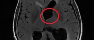
Diagnostics
Lateral dislocation of brain structures is detected during the study of Echo-EG (Echoencephalography). If the M-echo ultrasound signal reflected from the mid-brain is displaced by up to 4 mm, the displacement of the brain matter fragments is considered moderate. When the M-echo signal moves up to 10 mm, the shift is pronounced; more than 10 mm is critical. Other methods of instrumental research:
- MRI, CT. The most informative methods. Neuroimaging allows us to judge individual cases of dislocation and their combinations. The doctor receives information about the movement of the brain matter based on signs - incorrect location of the transparent septum and ventricular system (up to 4 mm - slight displacement, up to 9 mm - moderate, from 10 mm - pronounced), the shape and degree of enlargement of the cisterns.
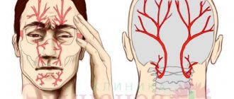
- Angiography. Deformation, compression, and displacement of blood vessels indicate movement of a fragment of the brain.
- Ultrasound diagnostics.
During a physical examination, the doctor determines the neurological status of the patient (the degree of depression of consciousness, the presence of cerebral and focal symptoms). At the same time, pulse and blood pressure are determined, and respiratory activity is assessed.
What's next?
For some reason, some patients tend to believe that the conclusion of an ES is already a sufficient reason to prescribe independent treatment, but this is certainly a big mistake.
A specialist diagnostician, of course, deciphers the echogram quite accurately, but the conclusion made on the basis of the examination results still includes only a preliminary diagnosis. A more accurate diagnosis can only be made by the attending physician who has a medical history and the results of other studies. You also need to take into account the fact that additional diagnostic methods and tests may be needed.
- Why is a study of the blood vessels of the brain and neck performed?
Thus, we must not forget in any case that after ES, you need to contact your doctor with its results, but definitely not draw up a treatment regimen for yourself.
As follows from the above, echoencephalography is a fairly reliable, informative, safe and certainly accessible method of studying the brain. There are no obstacles to its implementation and therefore, if there are specific indications, it is worth giving preference to this particular diagnostic method.
Treatment methods
Therapy is aimed at eliminating the cause of the pathological process - the primary disease (tumor, hemorrhage, infection, mechanical damage to the bones of the skull). Treatment of dislocation syndrome is carried out by a doctor in a comprehensive manner using conservative and surgical methods. If there are symptoms of medullary displacement, therapy is prescribed to remove excess fluid from the body (dehydration drugs) and against the development of edema (decongestants).
The patient is immediately prepared for surgery. Therapy with glucocorticoids makes it possible to stabilize the patient’s condition for some time (by reducing edema and lowering intracranial pressure values). With the development of life-threatening conditions, ventricular drainage and reclination (artificial, mechanical correction of the shift) of the brain are urgently performed.
In case of the lateral form of the pathology, external brain decompression is performed, followed by plastic surgery of the dura mater of the brain. If the pathological focus is localized in the posterior fossa of the skull, decompression craniotomy is performed, possibly followed by laminectomy of the upper vertebrae and opening of the dura mater.
Symptoms
This pathology is usually accompanied by a coma in the patient. However, loss of consciousness is not always observed.
For example, if the disorder was caused by a sudden process, swelling of the brain or an infectious lesion of the nervous system, the person may remain conscious.
Other factors can also lead to the development of brain dislocation, under the influence of which the abnormal process proceeds more slowly.
Typically it is characterized by the following symptoms:
- convulsions;
- temporary or permanent loss of vision;
- severe headaches;
- frequent vomiting and nausea.
Prognosis and prevention
In some cases, emergency medical attention is required. The progression of pathology is associated with a real threat of transition of a reversible syndrome to an irreversible stage. Possible consequences: deep coma, tachypnea (rapid shallow breathing), lack of pupillary response to a light stimulus, decerebrate rigidity (increased tone of the extensor muscles) type.
In extremely severe cases, a terminal (borderline with life) condition develops, which is characterized by hypoxia, acidosis, and acute general intoxication. To prevent pathology, doctors recommend avoiding head injuries, promptly treating infectious and tumor diseases of the central nervous system, and eliminating disturbances in the circulatory system.
Dislocation of the brain substance is a pathology that threatens life and health. Timely diagnosis and medical care will help avoid serious complications and death.
387